
Page 24 - Journal of Structural Heart Disease - Volume 1 Issue 2
P. 24
53
Meeting Abstracts
and April 2010 on a Philips Integris BH5000 biplane system. The study group underwent catheterization between April 2012 and October 2014 on either a Toshiba INFINIX-I CFI-BP or a Toshiba-I VFI-BP system. Each group contained 50 patients, who were matched by diagnosis, age, and weight. We recorded the dose area product (DAP) and fluo- roscopy time (FT) for each study. The effective radiation dose (ED) was determined using previously published conversion factors.
Results: The median age for the historical and study groups was 2.5 yr and 2.6yr, respectively. The two groups were equal in respect to body surface area (p=0.55) and FT (p=0.36). The median DAP in the study group was significantly lower than the historical group (5.8 Gy*cm^2 vs. 12 Gy*cm^2, p <0.001). Likewise, the median ED in the study group was significantly lower than the historical group (4.13 mSv vs. 10.84 mSv, p <0.001).
Conclusions: Advances in equipment technology in the cardiac cath- eterization laboratory can lead to significantly less radiation expo- sure, which is particularly important in the pediatric age group due to intrinsic properties of young tissue and stochastic effects over time. Institutions should aim, when feasible, to upgrade equipment in order to take advantage of this potential radiation dose reduction.
#0049
SUCCESSFUL PERCUTANEOUS RECANALIZATION OF A CHRONICALLY OCCLUDED IVC IN A CHILD
Emily Lawson1, Michael D. Seckeler1
1Department of Pediatrics, University of Arizona, Tucson, AZ, USA 2Department of Pediatrics (Cardiology), University of Arizona, Tucson, AZ, USA
We present the case of a 5 year-old girl with Scimitar syndrome with near-complete occlusion of her intrahepatic IVC with collateralization to hepatic veins for at least two years. She had had previous attempts at percutaneous management of her disease at another institution, which included device closure of a secundum ASD and PDA from a femoral approach. She was brought to the cath lab for potential reca- nalization of the IVC.
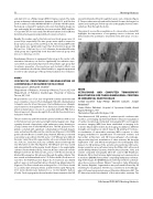
The procedure was performed under general anesthesia with access in the femoral vein and artery and right internal jugular vein. Angi- ography showed a hypoplastic right pulmonary artery draining to a large scimitar vein to the IVC-RA junction. The IVC appeared com- pletely occluded with significant collateralization through hepatic veins and entered the RA near the scimitar vein (Figure a). A 6-Fr MPA guide catheter (Boston Scientific Corp, Natwick, MA) was positioned as proximal as possible in the IVC and a 0.014” Whisper wire (Abbott Vascular, Santa Clara, CA), supported by a Corsair catheter (Asahi In- tecc USA, Inc, Santa Ana, CA), was carefully advanced through the true IVC lumen to the SVC (Figure b). The Whisper wire was snared and externalized from the jugular sheath (Figure c). An angled glide catheter (Terumo Medical Corp, Somerset, NJ) was advanced over the Whisper wire from the femoral vein, and a 0.035” Rosen wire (Cook Medical, Bloomington, IN) advanced through the glide catheter. This was snared and externalized and a 7-Fr Flexor sheath (Cook) was ad- vanced to the IVC-RA junction (Figure d). A small tract was dilated with a 6 mm x 3 cm Powerflex balloon (Cordis Corp, Miami Lakes, FL) (Figure e). The Flexor sheath was exchanged for a 10-Fr Flexor sheath to accommodate stent placement, and three telescoped Palmaz XL 3110 stents (Cordis) were delivered on a 12 mm OptaPro balloon (Cordis). Final angiography showed marked improvement in the IVC
size with diminished flow through the hepatic vein collaterals (Figure f ). The procedure was safe and well-tolerated. She was discharged on aspirin and clopidogrel. Follow-up echocardiography showed contin- ued phasic flow through the stents.
This patient’s successful recanalization of a chronically occluded IVC highlights the importance of recognizing venous occlusions and demonstrates the potential to treat them even after years of occlu- sion.
#0050
ULTRASOUND AND COMPUTED TOMOGRAPHY REGISTRATION FOR THREE-DIMENSIONAL PRINTING IN CONGENITAL HEART DISEASE
Jordan Gosnell1, Todd Pietila2, Bennett Samuel1, Joseph Vettukattil1
1Helen DeVos Children's Hospital of Spectrum Health, Grand Rapids, Michigan, USA
2Materialise, Plymouth, Michigan, USA
Three-dimensional (3D) printing of patient specific cardiovascular models is an emerging experimental field for enhanced visualization of cardiac morphology. Computed tomography (CT) and magnetic resonance imaging (MRI) have been established as imaging tools to derive 3D printable models. Each imaging modality has different strengths and weaknesses which impacts 3D printing: CT enhanc- es visualization of extracardiac anatomy; MRI is superior to other imaging modalities for quantification of ventricular volumes and myocardial architecture; and 3D transesophageal echocardiogra- phy provides the best visualization of valve anatomy. We describe registration of CT and 3D transesophageal echocardiography (TEE) datasets for 3D printing of the systemic atrioventricular (AV) valve. Image acquisition was performed using the Philips iE33 ultrasound system (Philips Medical Systems, Andover, Massachusetts, USA), and volume computed tomography (VCT) CT scanner (GE Healthcare, Waukesha, WI) in the Cartesian digital imaging and communication in medicine (DICOM) format. After completing multiplanar reformat- ting of the images, the DICOM datasets were imported into a dedicat- ed post-processing software (Mimics® Innovation Suite, Materialise NV, Leuven, Belgium). Segmentation was performed followed by 3D rendering for visualization. The file was converted to stereolithogra- phy (.stl) format and printed using a 3D printer (Materialise NV). The CT dataset provided visualization of the extracardiac anatomy. The
19th Annual PICS/AICS Meeting Abstracts
