
Page 46 - Journal of Structural Heart Disease - Volume 1 Issue 2
P. 46
75
Meeting Abstracts
cedure, compression of the puncture site in the neck for 15 minutes was enough to stop the bleeding. No hematoma was formed. The whole procedure lasted 50 minutes and the fluoroscopy time was 10 minutes.
Conclusion: We routinely use the femoral vein for closure of PDA. However, in the event of venous thrombosis or anatomical variations, we showed that the jugular vein can be safely used.
#0104
BAIL OUT RVOT STENTING IN A PREGNANT PATIENT WITH SEVERE ISOLATED INFUNDIBULAR STENOSIS Jayaranganath M, Usha MK, Srinivas KH
Sri Jayadeva Institute of Cardiovascular Sciences and Research, Banagalore, Karnataka, India
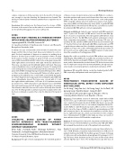
A twenty one year old lady with seven months of amenorrhea was diagnosed first time to have heart disease and referred to our hos- pital. She had complaints of dyspnea on exertion on walking short distance. She had cyanosis with room air saturation of 86%. She had harsh ejection systolic murmur of grade 3/6 left second and third space. ECG showed RVH with RV strain. Echocardiogram showed di- lated right atrium and ventricle with right ventricular dysfunction. There was a moderate size OS ASD with right to left shunt. There was severe infundibular stenosis with gradient of nearly 170 mmHg. Fetal echocardiogram was normal and fetal growth was adequate. She was planned for RVOT stenting keeping in my mind the hypoxemic effects on fetus and possibility of worsening RV failure in further weeks of pregnancy. Her abdomen was covered with a lead shield during the procedure. Right ventricle angiogram showed hypertrophied RV with severe infundibular stenosis.The lesion was crossed with 5 F Jud- kins right catheter with terumo 0.035 wire. A 2910 palmaz stent was mounted over a 14X40 atlas balloon and deployed across the RVOT. The right ventricle pressure fell down from 150 mmHg to 45 mmHg. The saturation after 24 hours was 92% and on follow up after 2 weeks was 95%. The fetal growth is satisfactory and she is symptom free. She has been continued on aspirin. She is due for delivery in first week of July.
Present angio Post stent
#0105
SUCCESSFUL DEVICE
DUCTUS ARTERIOSUS WITH TRANS-PULMONARY ECHOCARDIOGRAPHIC GUIDANCE WITHOUT CONTRAST ANGIOGRAPHY
Kenji Suda1, Yoshiyuki Kudo1, Hironaga Yoshimoto1, Yozo Teramachi2, Yoshiyuki Kagiyama1, Motofumi Iemura2 1Kurume University School Of Medicine, Kurume, Japan
2St. Mary's Hospital, Kurume, Japan
Background: Though contrast angiography is the standard guidance
of device closure of patent ductus arteriosus (DC-PDA), it is contra-in- dicated in patients with severe renal disease that often seen in senile patients. We have developed trans-pulmonary echocardiography (TPE) using ICE catheter placed in pulmonary arteries, main pulmo- nary artery (MPA) and left pulmonary artery (LPA), to guide DC-PDA (Cathet Cardiovasc Intervent 2015). We report 2 cases that successful- ly underwent DC-PDA without contrast angiography.
Materials and Methods: Subjects were 2 patients with PDA aged 48.5 and 71.2 years old. The sizes of PDA were 3.6 and 6.4 mm with Qp/ Qs of 1.7 and 1.9, respectively. The older patient suffered from renal dysfunction. Prior to the DC-PDA, both patients underwent contrast X-ray computed tomography to clarify the anatomy. TPE was ob- tained by the ICE catheter at MPA and LPA that was inserted through 2nd sheath at femoral vein. During the DC-PDA, we primarily used TPE to guide the procedure and also decided to minimize contrast angi- ography as long as we could comfortably perform the procedure. In the second case, we additionally used CARTO system to help under- stand the orientation of cutting plane of TPE.
Results: We could successfully close both PDA without any contrast angiography. TPE at MPA view and LPA view worked very well to de- termine the diameter and length of PDA, to monitor the device place- ment, and to determine the residual shunts. TPE did not increase the risk of complication except for transient arrhythmia, though new op- erator needs some learning time to understand orientation of TPE.
Conclusion: TPE using ICE catheter can be the standard guide for DC- PDA, especially adult patients with renal dysfunction.
#0106
PERCUTANEOUS TRANSCATHETER CLOSURE OF VENTRICULAR SEPTAL DEFECT USING AMPLATZER DUCT OCCLUDER
So Ick Jang1, Sang Yun Lee1, Jin Young Song2, Su Jin Park1, Mi Gyoung Song1, Hye Won Kwon1, Seong Ho Kim1
1Sejong General Hospital, Gyeonggi-do, Republic of Korea 2Samsung Medical Center, Seoul, Republic of Korea
Objectives: Transcatheter closure of ventricular septal defect (VSD) has become a good alternative treatment option to surgery. How- ever, complete atrioventricular block (CAVB) after device closure still remains a controversial issue. In our institute, Amplatzer Ductal Oc- cluder (ADO; St. Jude Medical Corporation, MN) is used to close the defect, since the shape of ADO is suitable for perimembranous VSD with aneurysm, due to the anatomical resemblance, and the device has no right ventricular disc, which is presumed to be the contribu- tion of development of CAVB. In this study, we show our results of transcatheter closure of VSD and the complications.
Methods: From Aug 2009 to Apr 2105, 39 patients underwent percu- taneous closure of perimembranous and muscular VSD with ADO . We reviewed the medical records of the patients retrospectively.
Results: 15 male and 24 female patients underwent transcatheter clo- sure of VSD. Of them, 24 patients had perimembranous VSDs and 15 patients had muscular VSDs. The median age was 6.6 years old (range 2.1-48.9 years old) and median body weight was 23 kg (range 8.0-75.7 kg). The mean shunt diameter was 4.6±1.7 mm and the mean shunt amount (Qp/Qs) was 1.6±0.3. The median fluoroscopy time was 23
CLOSURE OF
PATENT
19th Annual PICS/AICS Meeting Abstracts
