
Page 51 - Journal of Structural Heart Disease - Volume 1 Issue 2
P. 51
Meeting Abstracts
80
#0116
UNZIPPING OF SMALL DIAMETER STENTS AS A MANAGEMENT STRATEGY FOR NEONATAL COARCTATION OF THE AORTA IN A GROWING SWINE MODEL
Shyam Sathanandam1, Susheel Kumar1, Saradha Subramanian1, Ryan Sullivan1, Michael Perez1, David Zurakowski2, Christopher Knott-Craig1, Rush Waller1 1University of Tennessee, LeBonheur Children's Hospital, Memphis, TN, USA
2Harvard Medical School, Boston, MA, USA
Background: Surgical repair for critical coarctation of the aorta (COA) in the neonate is favoured over trans-catheter therapies. Small diam- eter stent (SDS) implantation may be effective in relieving the steno- sis acutely in this situation. However, the circumferential size of SDS implanted in a neonate does not adapt to the growth of the vessel leading to refractory stenosis in the future. If stents can be longitu- dinally fractured – unzipped, using angioplasty balloons, it can be re-dilated to the eventual adult vessel diameter as the child grows. The VeriFLEX and the Express stents were found to be most feasible and safe to unzip in a previously conducted in vivo experiment. The objective of this study was to determine the long term effects of using SDS to treat coarctation of the aorta in new-born piglets that would eventually be unzipped as the pig grows to adulthood.
Methods and Results: COA was surgically created in 10 new born pig- lets (Median weight 2.4 Kg). A poly-ethylene terephthalate band was applied in 5. Excision of half the aortic wall followed by application of a constricting suture was used in the other 5. A cardiac catheter- ization performed 4 weeks later (median weight 5.8 Kg), in 8 of the surviving piglets demonstrated significant angiographic stenosis and a peak systolic gradient of 34 ± 8 mmHg in both models. The stenosis measured 4 ± 1.5 mm. The COA was treated with implantation of a 4mm VeriFLEX stent in 5 piglets and a 6mm Express stent in 3 piglets. Repeat catheterization was performed when the piglets were 26 ± 6 Kg. Re-stenosis with a peak systolic gradient of 18 ± 12 mmHg was treated by dilation of the stents till they unzipped with no residual gradient. All stents unzipped without any complications. Four piglets were euthanized to determine the acute effects of stent unzipping. Re-catheterization was performed on the rest at 54 ± 8 Kg with fur- ther angioplasty or implantation of a large stent (Palmaz XL) to treat any residual stenosis. The medial dissection score on HP microscopy was 1 ± 0.8 acutely and 0.5 ± 0.5 in the chronic model.
Conclusions: It is feasible to implant a small diameter stent to treat coarctation of the aorta in new-born piglets. It is feasible and may be safe to unzip these stents without significant vessel wall injury. Unzip- ping of stents allows for future re-dilation or re-stenting with a large diameter stent preventing residual stenosis.
#0117
X-RAY FUSED WITH MRI (XFM) GUIDANCE OF TRANSCATHETER INTERVENTIONS IN CONGENITAL HEART DISEASE: PRELIMINARY RESULTS
Elena Grant1, Anthony Faranesh2, Laura Olivieri1, Joshua Kanter1, Russell Cross1, Michael Hansen2, Kendall O'Brien1, Karin Hamann1, Tina Ehtiati3, Robert Lederman2, Kanishka
Ratnayaka1
1Children’s National Medical Center, Washington, DC, USA 2National Heart Lung and Blood Institute, Bethesda, MD, USA 3Siemens Medical Solutions USA, Inc, Washington, DC, USA
Background: Interventional cardiologists are pursuing increasingly complex catheter based intervention. Fusion imaging provides soft tissue context that may simplify interventional procedures, reduce radiation exposure, and decrease contrast burden.
Methods: We prospectively recruited patients referred for transcath- eter device implantation. Radiation and contrast exposure for X-Ray Fused with MRI (XFM) cases was compared to intervention matched cases from our preceding 10 year institutional experience and Likert scale operator assessments of value were recorded.
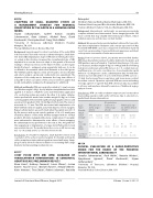
Results: From 11/2013 to 5/2015, 20 patients were enrolled. Cardiac MRI data indicated intervention should be deferred in 5 patients, and XFM guidance was performed in 15 patients [median age 10.6 years (2.5–62 years); median weight 47 kg (13–104 kg)] with the following indications: pulmonary artery (PA) stenosis (n = 7), conduit stenosis/ insufficiency (n = 3), aortic coarctation (n = 3), and ventricular septal defect (n = 2). Diagnostic cardiac catheterization data showed inter- vention was not indicated in 4 cases. Interventional XFM cases (n = 11) had shorter mean fluoroscopy times, and lower mean contrast dose than controls. Operators reported XFM was never misleading, “strongly agreed” or “agreed” that XFM soft tissue data was additive in all cases and may have allowed for omission of traditional angiog- raphy in 6 cases.
Conclusions: XFM can reduce radiation exposure and contrast dose by providing operators with useful soft tissue data in selected con- genital heart disease interventions.
Case Type Results: mean value (range)
PA balloon or stent angio- plasty
XFM n= 6
24 (13-44)
4.2 (2.6-5.6)
Control n = 142
42 (9-154)
5.9 (0.2-22)
XFM n= 3
11 (8-14)
2.6 (0.9-5.7)
Control n = 74
22 (7-100)
3.8 (0.8-11)
XFM n= 2
40 (21-59)
2.2 (1.0-3.3)
Control n = 26
43 (17-170)
7.0 (1.2-13)
Coarctation balloon or stent angio- plasty
Ventricular sep- tal defect device closure
Fluo- roscopy time (min)
Contrast (ml/kg)
#0118
CLINICAL EVALUATION OF A RADIO-PROTECTIVE CREAM FOR THE HANDS OF THE PEDIATRIC INTERVENTIONAL CARDIOLOGIST
Saradha Subramanian1, Rush Waller1, Natasha Winders1, Vijayakumar Agrawal1, David Zurakowski2, Shyam Sathanandam1
1University of Tennessee, LeBonheur Children's Hospital, Memphis, TN, USA
2Harvard Medical School, Boston, MA, USA
Journal of Structural Heart Disease, August 2015
Volume 1, Issue 2: 36-111
