
Page 56 - Journal of Structural Heart Disease - Volume 1 Issue 2
P. 56
85
Meeting Abstracts
Results: Twenty-one catheter interventions were performed in 10 patients. The median age was 30.9 years (range 20.1-63.0 years) and median weight 46.1kg (range 37.2-73.1kg). Only one patient, who had undergone lateral tunnel Fontan, was followed up from infant at our hospital. The remaining 9 patients were referred beyond adoles- cence. Stage of palliation at referral time was natural course in 2 pa- tients, Blalock-Taussig (BT) shunt in 4, bidirectional Glenn (BDG) in 1, original Fontan in 1 and Atrio-Pulmonary Connection Fontan in 1. An- atomical diagnosis was SRV in 4 patients, Tricuspid atresia in 3, DILV in 2 and DORV in 1. The CI prior BDG was one balloon angioplasty for shunt (5%), the CI between BDG and Fontan consisted of thirteen aortopulmonary collateral coil embolizations (61%) and one veno-ve- nous collateral coil embolization (5%), the CI after Fontan included two balloon angioplasty and one stent implantation for Fontan route (14%), two aortopulmonary collateral coil embolizations (10%), one veno-venous collateral coil embolization (5%). There were no major complications regarding catheter intervention in this study. One pa- tient who was natural course and two patients who had BT shunt at referral time had reached BDG, the remaining 7 patients had com- pleted extracardiac TCPC (including extracardiac TCPC conversion).
Conclusion: Catheter intervention plays an important role in the management of adult patients with single ventricle physiology.
#0129
THE USE OF DIGITAL SUBTRACTION 3-D ROTATIONAL ANGIOGRAPHY DURING CARDIAC CATHETERIZATION IN CHILDREN LESS THAN TWO YEARS OF AGE
Mario Briceno-Medina1, Emily Hayes1, Rush Waller1, Andrew Kuhls-Gilcrist2, Jason Johnson1, Lucas Elijovich1, Shyam Sathanandam1
1University of Tennessee, LeBonheur Children's Hospital, Memphis, TN, USA
2Toshiba America Medical Systems, Tustin, CA, USA
Background: Advantages of rotational angiography during cardiac catheterization include tomographic imaging, 3D road-mapping etc. Concerns over potentially higher contrast and radiation doses have limited its routine use in infants. We instituted a digital subtraction 3D rotational angiography (DS-3DRA) protocol for use in infants. The objective of this study was to compare radiation and contrast doses required for obtaining DS-3DRA with conventional digital 3DRA (DA- 3DRA) in children ≤ 2 years of age.
Methods: Radiation and contrast doses required for DS-3DRA was compared with age-, size- and diagnosis-matched historical controls that had DA-3DRA. Only children ≤ 2 years of age were included in the study. A 1:1 control matching was performed. Those patients that did not have matching controls were excluded from the study. The diagnostic quality and utility of these two modalities were scored by 4 qualified independent observers.
Results: The study (n=7) and control (n=7) groups were well matched for age (mean 14 vs. 15 months; p=0.239) and size (mean BSA 0.42 vs. 0.44 m2; p=0.103). The mean dose area product (DAP) to acquire DS- 3DRA was 34% higher than DA-3DRA (128 vs. 188 cGy.cm2; p=0.014). Similarly, the DS-3DRA air-Kerma, albeit small, was 47% higher than the DA-3DRA air-kerma (mean = 21.7 vs. 11.4 mGy; p<0.01). However, the contrast volume to acquire the best diagnostic quality DS-3DRA was 44% less than what was required for DA-3DRA (mean 1.02 vs. 1.81 mL/kg; p<0.001). The diagnostic quality and utility scores for the ro- tational angiography (86% vs. 84%; p=0.32), multi-planar reformation (84% vs. 88%; p=0.12), 3D reconstruction (79% vs. 86%; p=0.14), and 3D road-mapping (88% vs. 89%; p=0.36) were similar for both mo- dalities.
Conclusions: Digital subtraction rotational angiography can help reduce contrast volumes required to perform 3DRA in children ≤ 2 years of age. The radiation dose for DS-3DRA, albeit small, is a little over a third higher than for DA-3DRA. The diagnostic quality and utili- ty of DS-3DRA for infants with congenital heart diseases is equivalent to conventional 3DRA.
#0128
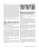
A NOVEL
APPROACH STEERABLE GUIDE CATHETER FOR A PATIENT WITH ATRIAL SEPTAL DEFECT AND IVC OCCLUSION
Teiji Akagi, Yoichi Takaya, Koji Nakagawa, Shunji Sano Okayama University, Okayama, Japan
A 11-year-old man was referred to our hospital for transcatheter clo- sure of atrial septal defect (ASD). Transesophageal echocardiography revealed an 11-mm secundum ASD with aortic rim deficiency. Be- cause he had a congenital occlusion of inferior vena cava,
To deliver a device via transjugular approach, we selected an Agilis steerable guide catheter (St. Jude Medical, St. Paul, MN), which was able to create an acute curve by the handle and to insert another 8 Fr sheath to load the device. Under fluoroscopy and transesopha- geal echocardiography guidance, the Agilis catheter was positioned across the defect into the left atrium, and then a 0.035 inch Amplatzer extrastiff guidewire (Cook, Indianapolis, IN) was advanced into the upper left pulmonary vein. A 13-mm Amplatzer Septal Occluder (St. Jude Medical, St. Paul, MN) was loaded into the sheath and advanced into the left atrium using the Agilis catheter. The position of the de- vice was confirmed by transesophageal echocardiography, and the device was released.
Because transjugular approach is difficult to maintain a pulmonary venous guidewire position and deploy the device stability, the proce- dure is very challenging.
The Agilis catheter is a steerable and adjustable curved guide cath- eter and has an insertion valve accommodating an 8.5 Fr catheter at the most proximal end to insert the device. This technique is useful in patients with IVC occlusion.
TECHNIQUE
USING TRANSJUGULAR
19th Annual PICS/AICS Meeting Abstracts
