
Page 61 - Journal of Structural Heart Disease - Volume 1 Issue 2
P. 61
Meeting Abstracts
90
#0140
TRANSCATHETER CLOSURE OF A MITRAL VALVE PARAVALVULAR LEAK IN A 7 MONTH OLD CHILD Ugonna Nwankwo, James Goldsmith, Sara Trucco, Jacqueline Kreutzer
Children's Hospital of Pittsburgh of UPMC, Pittsburgh, PA, USA
Objective: To describe successful percutaneous treatment of a para- valvular leak (PVL) in an infant with Shone’s complex and a supra-an- nular mechanical mitral valve (MV).
Background: Mitral valve replacement is a relatively rare procedure in young children. PVLs are a known complication of mitral valve re- placement. To our knowledge, this is the first report of a PVL treated percutaneously in a child this age.
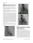
Case: The patient is a 7 m old, 6 kg, male with a history of Shone’s com- plex status post aortic arch reconstruction and balloon valvuloplasty of the MV. Due to severe mitral insufficiency, he underwent MV re- placement with a 16 mm Medtronic open pivot AP 360 mechanical aortic valve in the supra-annular mitral position at 5 months. He de- veloped a posterior PVL post-operatively with persistent hemolysis requiring repeated transfusions. Medical therapy with pentoxifylline failed. Thus, 2 m post-operatively, he was referred for percutaneous cardiac catheterization for PVL closure. Transesophageal echocardi- ography (TEE) identified a small-sized PVL between the mouth of the left atrial appendage and the left upper pulmonary vein. A trans-sep- tal approach was utilized. Using a Cobra catheter, a 0.018” floppy wire was advanced from the venous sheath through the leak and into the ascending aorta, where it was snared by a multisnare introduced from the arterial sheath. A 4mm Amplatzer vascular plug II was then positioned anterogradely and was successfully deployed in the tract. No significant leak was visualized on angiography or TEE and the child’s hemolysis subsided.
Conclusion: Device closure of mitral valve paravalvular leaks in a very small child is technically feasible, and may obviate repeat surgery.
Figure 1: Paravalvular leak (A), wire course (B), vascular plug after de- ployment (C).
#0141
A CASE REPORT: PERCUTANEOUS, TRANS-CATHETER CLOSURE OF THE CIRCUMFLEX TO LEFT VENTRICLE, CORONARY-CAMERAL FISTULA
Smita Mishra, S. Radhakrishnan
Jaypee Hospital, Sector 128 Noida,Up, India
Introduction: A 4 - year old male child presented with chest pain and failure to thrive. Clinically he had mild cardiomegaly, a loud P2 and a grade II/VI systolic murmur at left sternal border and apex. Radio- logically , he had unusual but mild localised protrusion at left heart border (Figure-1). Echocardiography was done on the IE33 Philips 3D Ultrasound System, which revealed a large coronary cameral fistula (CCF), arising from circumflex coronary artery just after its origin and terminating into LV. A Coronary cameral fistula (CCF) or coronary arte- rio-venous fistula(CAVF) is an uncommon type of shunt lesion due to presence of an abnormal fistulous tract between one of the branches of coronary artery and the cardiac chamber (CCF) or vascular struc- ture (CAVF). 1,2 A large fistula may present with chest pain, growth failure, congestive heart failure , arrhythmias or with life threatening
Journal of Structural Heart Disease, August 2015
Volume 1, Issue 2: 36-111
