
Page 24 - Journal of Structural Heart Disease Volume 3, Issue 3
P. 24
77
Meeting Abstracts
of 70-80%. He was awake with dusky oral mucosa. There was an equal chest expansion with no rales and wheezing. He had adynamic pre- cordium, point of maximal impulse at 4th ICS, S1 normal, S2 single with 3/6 systolic ejection murmur at LMCL. Liver was not palpable with no abdominal masses. There were full equal pulses with dusky nailbeds.
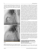
Figure 1.
Indication of Intervention: TOF is the most common form of cyanotic CHD. A 2002 meta-analysis of the incidence of CHD, which included 41 studies pertaining to TOF, suggested that the best estimate of
incidence would be 577 cases of TOF per million live births. Surgical options for management of symptomatic neonates and young infants with TOF include both complete repair and interim Blalock- Taussig (BT) shunt. However, there is signi cant peri-operative mor- bidity that includes prolonged mechanical ventilation, increased inotrope requirement and end organ dysfunction. The additional disadvantages include the need for ventriculotomy and higher risk of reoperation. Due to the increased demands of postoperative care coupled with these disadvantages, many centres are reluctant to attempt primary repair of TOF in infants less than three months of age. This is particularly true for centers in the developing world where the resources are limited. The alternative to corrective operation is palliation with BT shunt in very young infants, which is still advo- cated. The limitations of this procedure include the risk of distortion of branch pulmonary arteries in up to 15 to 20% and shunt occlusion in another 3 to 6%. In addition, there is signi cant postoperative mor- bidity and mortality following neonatal BT shunt. Balloon pulmonary valvotomy has been previously attempted in TOF as a palliative mea- sure. The right ventricular out ow tract (RVOT) obstruction in TOF is often at multiple levels: infundibulum, valve, annulus and, main and branch pulmonary arteries. Balloon pulmonary valvotomy can poten- tially o er reasonable interim palliation for infants with predominant valvar pulmonary stenosis (PS).
Intervention: Right heart catheterization was performed through a right femoral vein percutaneous puncture. A french 4 sheath was inserted and a french 4 pigtail catheter was manipulated under uoroscopic guidance into the IVC, RA, RV and LA through the pat- ent foramen ovale. Oximetry studies and pressure recordings were taken from selected vessel and chamber entered. RV angiogram was done using LAO, Cranial and lateral views showing opaci cation of the RV with passage of dye to the main pulmonary artery and to the aorta and its branches and PDA. Infundibular stenosis was noted on the right ventricular out ow tract. Con uent right and left pul- monary artery was also noted. The PDA was noted to be tubular and measured at 2 mm. PDA stenting was done using Omega Monorail 3x8mm. Exchange guidewire was inserted and placed in the periph- eral pulmonary artery. The catheter was removed with the guidewire in place and replaced by TMP Ped pulmonary valvotomy balloon catheter measuring 6 mm x 20 mm was inserted and in ated until the waist disappeared. Two in ations of the balloon catheter was done.
Learning Points of the Procedure: Balloon dilatation of the pulmonary valve is an e ective and safe palliation in tetralogy of Fallot. It promotes growth of the pulmonary vascular tree, reducing the need for trans-an- nular patching and is recommended in symptomatic infants of very young age, with a small pulmonary annulus (Z value below - 4 SD) and associated cardiac anomaly. (Eur Heart J 1998; 19: 595–600). The patient had improved oxygenation at 80-90 % discharged after with regular fol- low up awaiting total correction.
CLOSURE OF VENTRICULAR SEPTAL RUPTURE USING VENTRICULAR SEPTAL OCCLUDER AFTER MYOCARDIAL INFARCTION
Eloisa Victoria Claveria-Barrion, Jean Antonio Villareal Philippine Heart Center, Manila, Philippines
History and Physical: This is a case of a 64 years old female, newly diagnosed with diabetes mellitus, who came in due to chest pain.
Hijazi, Z
20th Annual PICS/AICS Meeting Abstracts
