
Page 25 - Journal of Structural Heart Disease Volume 3, Issue 3
P. 25
Meeting Abstracts
78
Two months prior to admission, patient had sudden onset of severe chest pain. She was subsequently admitted at the National Kidney and Transplant Institute and managed as a case of Acute Coronary Syndrome. During this admission, the patient had episode shortness of breath associated with orthopnea and exertional dyspnea. Two- dimensional echocardiography was done which revealed ejection function of 62 – 69 %, concentric left ventricular hypertrophy with hypokinesia of the left ventricular apex with suspicious ow across the muscular interventricular septum indicating a ventricular sep- tal rupture shunt. The patient was stabilized and was discharged improved.
Five days prior to admission, she was noted to have progressive bipedal edema. She was then readmitted at the NKTI. One day prior to admission, she was noted to have shortness of breath along with epigastric pain and vomiting. The patient was transferred to our insti- tution for intervention.
On physical examination, the patient had stable vital signs, not in distress and ambulatory. Neck veins were distended. On chest examination, there was no lagging and retractions but with crackles on bilateral mid to lower lung eld. The patient had adynamic pre- cordium, point of maximal impulse at the 5th intercostal space left mid clavicular line, with thrill at the left parasternal area, no heave, S1 normal, S2 split, normal rate, regular rhythm, grade 4/6 holosystolic murmur over left parasternal and apical area. The abdomen was soft, not distended, no ascites but with palpable liver edge 3 cm below subcostal margin. The lower extremities showed grade II pitting bipedal edema with full pulses.
The assessment on admission was Atherosclerotic Heart Disease, Coronary artery disease, s/p Acute coronary syndrome (March 2016), Ventricular septal rupture, Congestive Heart failure, NYHA Functional classi cation II-III, Diabetes Mellitus type 2.
On admission, the patient was worked-up. She underwent coronary angiography with noted one vessel disease (LAD). On LV angiogram, contrast injection showed passage of dye from LV to RV through the muscular part of the interventricular septum. On the 6th day, the patient underwent ventricular septal rupture device closure using VSD occluder size 17/10. The patient was able to tolerate the proce- dure well. On the 18th hospital day, the patient then underwent PCI.
Indication of Intervention: The indication for closure of an interven- tricular septal defect after acute myocardial infarction causing hemo- dynamic compromise with evidence of loud holosystolic murmur and left ventricular dysfunction with lower extremities edema is war- ranted in our patient. The prognosis of post – AMI VSD is very poor, with mortality rates as high as 50 % at 1 week and 90 % at 2 months with conservative medical management. The patient had a history of AMI 2 months prior to admission and surgery is technically di cult owing to the myocardial tissue being soft and friable. Percutaneous closure device closure is a viable option in chronic period in patients with co morbidities and whose septal anatomy is favourable to device placement. The patient presented with ventricular dysfunction, with history of diabetes which put her to a high risk candidate for surgery. The septal anatomy of the ventricular rupture of the patient was at the muscular area, which is the only area recommended by the American Heart Association for device closure.
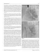
Figure 1.
Intervention: The patient underwent device closure of the muscular ventricular septal rupture on the 6th hospital day of admission. Left heart catheterization was performed via the right femoral artery per- cutaneous puncture and a French 6 sheath was inserted. A French 6 pigtail catheter was manipulated under uoroscopic guidance into the descending aorta, ascending aorta and to the LV. LV angi- ography at LAO 35, cranial 35 showed a muscular ventricular septal defect. Right heart catheterization was performed through a right femoral vein percutaneous puncture. A French 6 sheath was inserted via the right femoral vein. A French 6 multi-snare catheter was then inserted and manipulated under uoroscopic guidance into the IVC, RA and RV. An arteriovenous guide wire splint was then created. The
Journal of Structural Heart Disease, June 2017
Volume 3, Issue 3:73-95
