
Page 26 - Journal of Structural Heart Disease Volume 3, Issue 3
P. 26
79
Meeting Abstracts
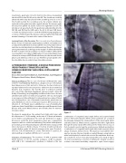
long Terumo guide wire 032 x 260 mm from the LV was manipulated thru the VSD to the RV, RA and to the IVC. The sheath was carefully advanced until its tip was placed in the ascending aorta. As soon as the sheath reached the ascending aorta, the arterial catheter was replaced via the guide wire with a pigtail catheter. The terumo guide wire was then pulled out by the snare to the right femoral vein. The introducer set was attached to the guide wire and pulled back to the IVC, RA, and RV thru the VSD and to the LV. A 6/8 mm VSD device occluder was then placed to occlude the VSD. Cineangiography post occlusion of VSD showed the device positioned within the VSD, with minimal shunting of contrast in the center of the device.
Learning Points of the Procedure: This is a novel case of percutaneous device closure of ventricular septal rupture post-MI in our institution. In a study done by Demkow et al, transcatheter closure has improved sur- vival rates in selected patients in suitable anatomy. One of the challenges among interventional cardiologist is the margins of defect wherein the borders on may be necrotic and the poor clinical condition of the patient on presentation. According to Bialkowski et al, procedure failures were observed in acute post-MI VSD and satisfactory in subacute and chronic phase cases which was seen in our case. Therefore, proper patient selec- tion should be done in order to have a favorable outcome.
LUTEMBACHER SYNDROME: A DOUBLE PROCEDURE (PERCUTANEOUS TRANSEPTAL MITRAL COMMISSURROTOMY AND ATRIAL SEPTAL DEFECT CLOSURE
Eloisa Victoria Claveria-Barrion, Jean Villareal, Juan Reganion
Philippine Heart Center, Manila, Philippines
History and Physical: This is a case of a 45 year old female who came in due to easy fatigability. Patient was a diagnosed case of rheumatic heart disease since 1980 but lost to follow up. She had a history of repeated admission for one year prior to admission due to exertional dyspnea and congestion. Two months prior to admission patient was seen at the outpatient department and was noted to have dif- culty of breathing, easy fatigability and bibasal rales hence she was admitted and started on medical management. She was discharged in an improved state with plan for interventional procedure. Patient was discharged as a case of rheumatic heart disease, mitral stenosis, atrial brillation in chronic ventricular response, NY functional clas- si cation II – III. Patient was re-admitted as a case of Lutembacher Syndrome, Atrial Septal Defect secundum type and Rheumatic Heart Disease with severe Mitral Stenosis two months after for interven- tional procedure.
Figure 1.
combination of congenital atrial septal defect and acquired mitral stenosis. The haemodynamic e ects of this syndrome are a result of the interplay between the relative e ects of the atrial septal defect and mitral stenosis. Mitral stenosis augments the left to right shunt through the atrial septal defect. Because the mitral stenosis was, in fact, rheumatic in aetiology, the syndrome was de ned eventually as a combination of congenital atrial septal defect and acquired, almost always rheumatic, mitral stenosis. Percutaneous transcatheter ther- apy has become the most widely accepted therapy, using balloon mitral valvuloplasty for mitral stenosis and the amplatzer atrial sep- tal occluder for closure of an atrial septal defect. In a study of Uguro SU, et al , percutaneous correction is preferred to surgical correction as there is decreased morbidity compared to open- heart surgery.
On physical examination, the patient had stable vital signs with blood pressure of 110/60 mmHg, cardiac rate of 76 beats per minute, not in distress and ambulatory. No neck vein distention nor palpa- ble masses noted. On chest examination, there was a symmetrical expansion with no crackles and wheezing. The patient had adynamic precordium, point of maximal impulse at the 5th intercostal space left intercostal space, no thrill, no heave, S1 normal, S2 split, normal rate, regular rhythm, grade 3/6 diastolic murmur at the apex. The abdo- men was soft, not distended, no ascites. There were no cyanosis, no edema with good capillary re ll time at < 3 seconds.
Indication of Intervention: The incidence of ASD in patients with mitral stenosis is 0.6-0.7% and the incidence of MS in patients with ASD is 4%. Lutembacher’s syndrome is de ned as the rare
Hijazi, Z
20th Annual PICS/AICS Meeting Abstracts
