
Page 31 - Journal of Structural Heart Disease Volume 3, Issue 3
P. 31
Meeting Abstracts
84
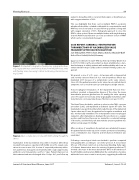
Figure 1. Computed Tomography of pulmonary angiography show- ing a large pulmonary arteriovenous stula from left pulmonary artery with feeding artery measuring 14.5mm and draining channel measur- ing 12.5mm.
Figure 2. Duct occlude device in situ with mild foaming through the device.
examination chest was bilateral clear, with no murmur. Chest X-ray revealed homogenous opacity in left middle zone. His oxygen satu- ration at room air was 93%. In view of suspected pulmonary infarc- tion patient was managed conservatively with cough suppressants, antibiotics and tranexaminic acid for 7 days. Patient improved on this treatment and was discharged after 10 days. At 1 year follow up
patient is doing ne with no recurrent hemoptysis or breathlessness with oxygen saturation of 93%.
This case highlights that there can be multiple PAVF in a patient, whether all should be occluded is debatable. In our patient the small PAVF was not closed and at one year follow up patient is doing well with oxygen saturation of 93%. Retrograde approach to close the PAVF is other option if there are multiple feeders with single draining channel. Pulmonary infarction can occur post device closure of PAVF which can be conservatively managed.
CASE REPORT: CEREBRAL STENTRETRIEVER THROMBECTOMY OF AN EMBOLIZED VALVE FRAGMENT AFTER VALVE IN VALVE TAVI
Jan-Erik Guelker, Peter Schott, Marcus Katoh, Alexander Bufe Helios Clinic Krefeld, Krefeld, Germany
Case: Successful valve in valve TAVI was rst reported by Grube et al in 2007 [1]. Until now the procedure has been established as a stan- dard technique in elderly patients with multimorbidity who bear an enhanced risk for major cardiac adverse events following cardiac sur- gery [2].
We present a case of a 78 - year - old woman with a degenerated and severely stenosed Hancock II 21 mm bioprothesis which was implanted 2007 because of a symptomatic aortic valve stenosis. Since 2014 the patient presented aortic valve stenosis related clinical symptoms such as recurrent syncope and dyspnea at light exercise.
Transoesophageal reevaluation of the implanted Hancock II bio- prothesis revealed a degenerative disease of the valve, the mean transvalvular pressure gradient was 50 mmHg, the valve opening area 0.50 square cm. Coronary artery angiography excluded an addi- tional stenotic coronary artery disease. The Euroscore was 18.05 %.
The Heart Team decided to perform a valve in valve TAVI, a proven procedure [3,4,5], and implanted an Edwards Sapien XT valve. The implantation was performed transfemorally, the size of the implanted Sapien valve was 21 mm; the patient had a regular anaesthesia and was mechanically ventilated. The intraoperative transoesophageal evaluation after implantation displayed the prosthesis in a regular position and function; there was no mismatch, no relevant leakage of the valve, and the nal peak transvalvular pressure gradient was within normal range (19 mmHg).
After cessation of anaesthesia the patient was extubated. However the patient failed to wake up and to resume spontaneous breathing. Soon a reintubation was required, and mechanical ventilation was reinstalled.
Following a neurological check we decided to perform computed tomography (CT) according to our stroke protocol. A noncontrast CT-scan of the brain ruled out an intracranial bleeding. Early signs of ischemic demarcation were also missing. However perfusion CT showed a decrease in the mean transit time (MTT) in the territory of the left middle cerebral artery (MCA) while the cerebral blood volume within the lesion was normal in most parts, suggesting the a ected
Journal of Structural Heart Disease, June 2017
Volume 3, Issue 3:73-95
