Original Scientific Article
 Download PDF (2.18 MB)
Journal of Structural Heart Disease, April 2016, Volume 2, Issue 2:44-45
DOI: 10.12945/j.jshd.2016.015.14
Transcatheter Mitral Valve Replacement: Update on New Devices
Matthew J. Gillespie, MD1,2*, Robert C. Gorman, MD2,3, Joseph H. Gorman III, MD2,3
1 The Children’s Hospital of Philadelphia, Division of Cardiology, Philadelphia, PA, USA
2 Gorman Cardiovascular Research Group, Department of Surgery, Philadelphia, PA, USA
3 Perelman School of Medicine, University of Pennsylvania, Philadelphia, PA, USA
Abstract
Transcatheter Mitral valve replacement represents the next frontier in cardiac valve therapy. This review article details the authors' experience thus far in the development of a novel catheter-based mitral valve replacement device, and also highlights the preliminary experiences of other new technologies.
Media
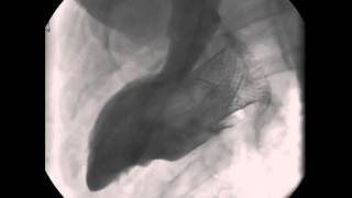
Video 1
An aortagram following Melody VIR, and importantly there is no aortic valve insufficiency.
Video 2

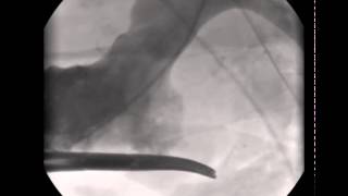
A Melody valve compressed onto a delivery balloon being advanced transvenouslyacross the atrial septume and subsequently withing the “incomplete annuloplastyring”.
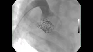
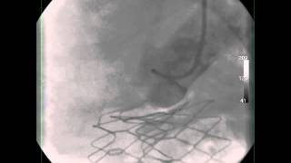
Video 3
The Melody valve securely seated within the incomplete annuloplasty ring. There is excellent systolic fuinction and no LVOT obstruction.
Video 4

This movie shows that there is no aortic valve insufficiency following Melody-in-incomplete-ring procedure. This is a very importatnt finding, because up until this point, it was generally believed that absence of a ring along the anterior leaflet would result in deformation of the aortic valve due to the mitral-to-aortic valve fibrous continuity. These experiments proved that this was indeed not true.
Video 5

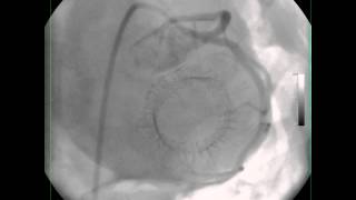
Color Doppler interrogation shows mild central MR, and no perivalvular leak.This video show typical results following Melody valve-in-ring mitral implantation.
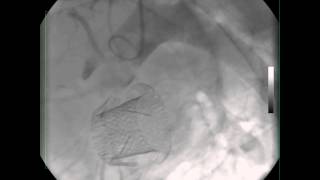
Video 6

A 2d image of the Melody valve and it’s relationship to the aortic valve. This video show typical results following Melody valve-in-ring mitral implantation.
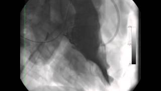
Video 7
LV gram post SMV replacement.
Video 8
Aortagram post SMV replacement.
Video 9
Video 10
This movie depicts a baseline left ventriculogram in a porcine model.
Video 11

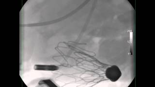
The sequence of valve deployment is initiated, beginning by withdrawing the outer sheath to allow the ventricular arms of the device to blossom. Once the ventricular side is opened, the device is withdraw towards the left atrium. This “traps” the anterior and posterior leaflets of the native mitral valve between the body of the SMV device and the ventricular arms. This is important for secure anchoring in the mitral position.
Video 12
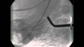
Once the ventricular end of the device is set, the outer sheath is fully withdrawn, allowing the atrial side of the device to open on the LA side of the mitral annulus.
Video 13
Video 14
Video 15

Video 16

Cite this article as: Gillespie MJ, Gorman RC, Gorman III JH. Transcatheter Mitral Valve Replacement: Update on New Devices. Structural Heart Disease 2016;2(2):44-45. DOI: 10.12945/j.jshd.2016.015.14
All comments will be screened and reviewed before posting. Statements, opinions, and results of studies published in Journal of Structural Heart Disease are those of the authors and do not reflect the policy or position of The Journal and Science International and the Editorial Board and provides no warranty as to their accuracy or reliability. Material is copyrighted and owned by Science International and cannot be used without expressed permission.
Original Scientific Article
Journal of Structural Heart Disease, April 2016, Volume 2, Issue 2:44-45
DOI: 10.12945/j.jshd.2016.015.14
Transcatheter Mitral Valve Replacement: Update on New Devices
Matthew J. Gillespie, MD1,2*, Robert C. Gorman, MD2,3, Joseph H. Gorman III, MD2,3
1 The Children’s Hospital of Philadelphia, Division of Cardiology, Philadelphia, PA, USA
2 Gorman Cardiovascular Research Group, Department of Surgery, Philadelphia, PA, USA
3 Perelman School of Medicine, University of Pennsylvania, Philadelphia, PA, USA
Abstract
Transcatheter Mitral valve replacement represents the next frontier in cardiac valve therapy. This review article details the authors' experience thus far in the development of a novel catheter-based mitral valve replacement device, and also highlights the preliminary experiences of other new technologies.
Media
Video 1
Video 2
Video 3
Video 4
Video 5
Video 6
Video 7
Video 8
Video 9
Video 10
Video 11
Video 12
Video 13
Video 14
Video 15
Video 16
PDF
Mobile-ready Flipbook
Cite this article as: Gillespie MJ, Gorman RC, Gorman III JH. Transcatheter Mitral Valve Replacement: Update on New Devices. Structural Heart Disease 2016;2(2):44-45. DOI: 10.12945/j.jshd.2016.015.14
You must be registered and logged in to leave comments.
There have been no comments posted yet
Ask a question (publicly)
Board