
Page 28 - Journal of Structural Heart Disease - Volume 1 Issue 1
P. 28
Original Research Article
22
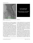
Figure 2. Micropuncture contrast injection after fluoro guided femoral access to ensure sheath insertion in a non-calcified seg- ment of the common femoral artery.
monary wedge pressures, or refractory symptomat- ic heart failure, may be stabilized enough by BAV to make non-cardiac surgery more tolerable for both the patient and the Anesthesia Team [7].
Recently, the major use of BAV has been for pre- dilatation during TAVR procedures. While the need for predilatation can be argued for some patient groups and for many average TAVR procedures, another utility of BAV for TAVR is to help with valve annulus sizing or choice of prosthesis size [8]. When predila- tation with a balloon matched to the expected annu- lus size yields a sealing of the annulus evidenced by contrast injection during the balloon inflation, great- er confidence can be found for a TAVR valve with the frame corresponding to the balloon size. Converse- ly, when the balloon will not lock in the valve, and a contrast injection during balloon inflation results in clear contrast regurgitation into the left ventricle, the balloon presents an undersized diameter for a given valve annulus.
Special care must be taken to be sure that the ful- Structural Heart Disease, May 2015
Figure 2 Video.
ly inflated predilatation balloon size measurement is accurate. Many balloons are manufactured with some variability in the technical specification. In ad- dition, hand inflation using a large syringe typically results in under-filling of the balloon, and it is likely in that setting that the balloon will not achieve its nominal diameter. Two methods to insure correct and full inflation include either use of a volume driv- en inflation with an inflation device, or the addition of a side syringe and a high pressure stopcock to the larger hand inflation syringe.
BAV can be used as an aid to sizing for TAVR [9]. It is not uncommon to have ambiguity for the selection of TAVR device size, even with CT annulus measure- ments. This problem results when patients are truly on the borderline between valve prosthesis sizes based simply on measurements, and is exacerbated by the challenges of heavily calcified leaflets or left ventric- ular outflow tract, and the underappreciated prob- lem of suboptimal CT scan images for analysis. One method favored by some operators to help resolve
Volume 1, Issue 1: 20-32
