
Page 36 - Journal of Structural Heart Disease Volume 1, Issue 3
P. 36
Original Scientific Article 142
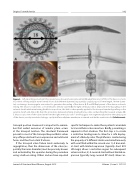
Figure 3. Self expanding valved stent for percutaneous tricuspid valve replacement (Boudjemline et al. réf 36). A. The device consists in a nitinol self-expandable stent formed of two disks (40-mm diameter) separated by a tubular part (15-mm length, 18-mm diame- ter) containing a bovine jugular vein valved. to guarantee the sealing of the device. B, C and D. Deployment of the device on bench testing. The device is loaded into a « homemade » delivery system (B), the right ventricular disk is deployed in the by pulling on the external sheath while maintaining the dilator in position; this disk is subsequently applied to the tricuspid annulus by pulling on the external sheath and dilator (C), then the atrial disk is delivered similarly, making the two disks sandwiching the tricuspid annulus (D). E. Macroscopic view of the valved stent from the right ventricular side. * bovine jugular vein segment sutured into the tubular part of the device; avoid paravalvular leakage, a polytetrafluoroethylene membrane is sutured outside the ventricular disk (black arrow).
tricuspid position. However it is important to remem- ber that widest excursion of annular plane occurs at the tricuspid location. The structural framework present in most of the tricuspid bioprosthetic valves may offer protection from compressive and rotational forces and thus from stent fractures.
If the tricuspid valve failure leads exclusively to regurgitation, then the dimensions of the valve (es- pecially the inner diameter) must be precisely known and evaluated by the operator during the procedure using a balloon sizing. Others and we have reported
specific techniques to make these patients amenable to transcatheter valve insertion. Briefly, presenting is required in that situation. The first step is to create a restrictive landing zone to allow for a safe deploy- ment of a Melody valve. The platform is created using the property of 2 different stents used simultaneously with one fitted within the second one: 1.) A bare met- al stent with limited expansion (typically short EV3 LD mega) allows a restrictive region for subsequent valve insertion; 2.) A covered stent without limited ex- pansion (typically long covered CP stent) allows an-
Journal of Structural Heart Disease, August 2015
Volume 1, Issue 3: 137-151
