
Page 15 - Journal of Structural Heart Disease Volume 2, Issue 1
P. 15
9 Review Article
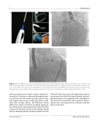
Figure 5. Panel A. LARIAT image courtesy of SentreHeart, Inc. Panel B. This left anterior oblique projection demonstrates epicardial and endocardial magnet wires connected at the tip of the left atrial appendage (LAA). An additional wire for maintained pericardial access is also seen. Panel C. This right anterior oblique uoroscopy image demonstrates the LARIAT snare being placed over the appendage while the endocardial and epicardial magnet wires function as a rail. Once over the base of the LAA, the snare will be closed.
cu around the face for direct contact with the en- docardium. The device is delivered through the fem- oral vein into the left atrium via trans-septal access and requires uoroscopy and TEE guidance as with other LAA occluder devices. The Wavecrest device di ers from others in that the occluding atraumatic face of the device is deployed rst into the LAA at the ostium and advanced outside of the delivery sheath without requiring delivery sheath placement into the
LAA itself. Once in position, the deployment anchors are advanced into the LAA body. Currently, dual anti- platelet therapy is recommended after implantation. This device has received CE mark approval. There is limited data concerning clinical outcomes with this device at this time.
Sánchez, J.M. et al.
Where Do We Stand Now?
