
Page 13 - Journal of Structural Heart Disease Volume 2, Issue 5
P. 13
Original Research Article
214
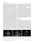
Catheterization was performed in a single-plane catheterization suite with a C-arm under general anesthesia with transesophageal echocardiography (TEE) guidance. TEE con rmed the presence of a 6 mm ruptured right sinus of Valsalva aneurysm with continuous ow into the RVOT (Figure 1:Panel A and B). These ndings were con rmed with ascending aortic angiography (Figure 2:Panel A). The mean pulmonary artery pressure was 26 mmHg, with right ventricular systolic pressure of 42 mmHg and ascending aortic pressures of 91/38. The ratio of total pulmonary to total systemic blood ow (Qp:Qs) measured 4.2:1 but was calculated on 100% oxygen as calculation in room air was not feasible. The defect was easily crossed with a 5-French (Fr) Judkins Right catheter and an exchange-length Glide wire with advancement of the JR4 into the distal right lower pulmonary artery. A 0.035” Amplatzer Extra-Sti wire (St. Jude Medical, St Paul, MN, USA) was positioned in the right pulmonary artery, and the JR4 was exchanged for a 5Fr guiding sheath (Figure 2:Panel B). A 6:6 Amplatzer Ductal Occluder (ADO) II (St. Jude Medical) was then advanced across the defect, the distal disk was partially deployed, and the sheath was retraced slowly from the main pulmonary artery. As the device crossed the pulmonary valve, the distal disk of the device engaged the right ventricular aspect of the defect, and the waist was deployed within the neck of the aneurysm and nally the proximal disk in the aortic root. TEE con rmed good device positioning (Figure 1:Panel C), but it was unclear if there was a small persistent leak. There was no impingement on the aortic or pulmonary valves. It was decided to release the device because there was some tension
from the delivery cable distorting the aortic end of the device away from the root. Following release, the device position was more satisfactory, and the nal aortic root angiogram con rmed good position with no residual leak (Figure 2:Panel C).
The patient recovered well from the procedure with no procedural complications. Predischarge echocardiogram the following day demonstrated a good device position, but it initially appeared that there may be a small residual leak. Further assessment revealed that there was a small perimembranous ventricular septal defect (VSD) just beneath the right coronary cusp of the aortic valve that was not initially noticed due to its proximity to the more prominent jet from the ruptured sinus of Valsalva aneurysm. The ow pattern was systolic with no diastolic component and a peak estimated systolic gradient of 82 mmHg. There was no RVOT obstruction or increase in the degree of mild aortic incompetence.
Discussion
Although surgical repair of ruptured sinus of Valsalva aneurysm has been extensively described [2], reports of transcatheter closure are sparse [3-9]. The largest study to date reported 18 successful cases from 20 attempts with a median age of 23 years (17–52 years) [3]. The majority of patients were symptomatic, and few had associated lesions such as VSDs, which were reported in 41% of 725 patients from a large systematic review [2]. In all patients, the defects were closed from the venous side using ADOs 2–4 mm larger than the aortic end of the defect. The ADO sizes ranged from 8/6 to 16/14 mm. Thirteen
Figure 1. Series of transthoracic echocardiogram images demonstrating the color ow across the ruptured sinus of Valsalva aneurysm from the right coronary sinus into the right ventricular out ow tract in the long (Panel A) and short (Panel B) axes. (Panel C ). The Amplatzer Ductal Occluder II is in a good position across the ruptured aneurysm following release.
Journal of Structural Heart Disease, October 2016 Volume 2, Issue 5:213-216
