
Page 24 - Journal of Structural Heart Disease Volume 2, Issue 5
P. 24
225
Introduction
Kawasaki disease (KD) is an acute febrile disease of unknown origin, characterized by systemic vascular in- ammation involving small and medium arteries, with predilection for coronary arteries. Since the rst report in 1967 from Japan [1], KD has become the most com- mon form of pediatric systemic vasculitis. The clinical presentation of KD is variable but classically consists of 5 days of fever, accompanied by nonpurulent conjunc- tivitis, rash on the trunk, erythema of the lips or oral cavity, erythema of hands or feet, and cervical lymph- adenopathy. An atypical presentation is di cult to rec- ognize; it may lead to delayed treatment and is thus associated with a higher incidence of coronary aneurysms (CAAs). Immediate treatment with immu- noglobulins reduces the incidence of coronary artery aneurysms in children from 25% to only 5% [1].
Nowadays, CAAs following KD are the leading cause of acquired heart disease in children and young adults in western countries. Coronary artery abnor- malities include persistent aneurysms with the risk of thrombosis and progressive stenosis, with or without the development of extensive collateral circulation. CAAs can persist, progress to stenosis, and lead to acute myocardial infarction [2]. There are few reports of coronary interventions after KD performed in young patients [3]; additionally, due to the highly variable anatomy of the lesions of the coronary arter- ies after KD, no con rmed standardized treatment guidelines for this particular patient group exist.
Here we report a case of successful and complete revascularization of thrombosed giant aneurysms of the right coronary artery (RCA) including dilatation of severe stenosis in a patient after atypical KD.
Case Presentation
A 10-year-old male child was transferred to our hospital with the presumed diagnosis of acute myo- cardial infarction. He su ered from two episodes of ventricular brillation that were successfully treated by resuscitation and de brillation. The electrocardio- gram (ECG) showed ST-segment elevation in V1 to V4 and was consistent with myocardial ischemia. The laboratory studies showed an elevated troponin I (11,232 pg/ml, Ref. 0–26; Figure 1). All other
Case Report
laboratory parameters were normal, including in ammatory markers. Transthoracic echocardiogra- phy showed dilatation of the central coronary arteries
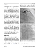
Figure 1. Panel A. Selective coronary angiography of the left and right coronary arteries. Panel B. The left coronary artery shows moderateaneurysmaticdilatationoftheleftanteriordescend- ing artery. The right coronary artery is completely occluded (both in strict a. p. 0° projection).
Meyer, Z. et al.
Revascularization after Atypical Kawasaki Disease
