
Page 8 - Journal of Structural Heart Disease Volume 2, Issue 5
P. 8
209
Original Research Article
resection, or even right pneumonectomy [5]. Successful attempts of interventional therapy of scimitar syndrome include transcatheter emboliza- tion of anomalous systemic arterial supply with inter- ventional atrial septal defect closure [6] and rerouting of anomalous venous drainage to the left atrium [7].
Case Report
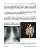
We present a 28-year-old female who was diag- nosed with right-sided PAPVR as a variant of scimitar syndrome. The patient had been a preterm infant born at 26 weeks of gestation. She developed bron- chopulmonary dysplasia and su ered from recurrent respiratory tract infections in infancy and early child- hood. Her psychomotor and physical development were unremarkable. At the age of 28 years, the patient presented to our unit with exertional dysp- nea. On chest x-ray, a tubular structure in the right cardiophrenic angle compatible with the typical ra- diologic sign of scimitar syndrome was evident (Figure 1). Subsequent MRI revealed anomalous return of the right lower pulmonary vein into the IVC. The scimitar vein was slightly stenotic proximal
Figure 1. Chest x-ray with a classic “scimitar sign” resulting from a right pulmonary vein coursing to the right cardiophrenic angle (arrows).
toitsdrainageintothedilatedIVC.Furthermore,all right-sidedpulmonaryveinswereconnectedviathe scimitar vein and drained not only into the IVC but also into the left atrium, thus representing a rare variant of scimitar syndrome. The right pulmonary veins had a more tortuous appearance than usual, but no discrete stenosis was noted (Figure 2). The right ventricle was enlarged due to volume overload, and systolic function was within normal limits. The pulmonary to systemic ow (Qp:Qs) ratio, as assessed by MRI, was 1.4:1.
Cardiac catheterization was performed with the intention of interventional closure of the aberrant pulmonary vein. Levophase after right pulmonary angiography con rmed dual connection of all right pulmonary veins via the scimitar vein to the IVC, as well as to the left atrium (Figure 3A). The left-to-right shunt ratio (according to Fick) was calculated to be 1.6:1. The patient’s right ventricular systolic pressure
Figure 2. Three-dimensional reconstruction of cardiac MRI (posterior-anterior view). The anomalous drainage of the lower pulmonary right vein into the IVC with slight stenosis of the vessel at the diaphragm is evident. In addition, all three right pulmonary veins are connected to the left atrium. AO = descending aorta; IVC = inferior vena cava; LA = left atrium; RPA = right pulmonary artery; * pulmonary veins; + scimitar vein; arrows, additional vein connecting the lower and middle right pulmonary vein.
Dieks, J-K. et al.
Interventional Treatment for Scimitar Syndrome
