
Page 45 - Journal of Structural Heart Disease Volume 2, Issue 6
P. 45
Meeting Abstracts
272
Results: A total of 58 patients with single ventricle had 72 stents implanted in the LPA (n=41, 57%), RPA (n=7, 10%), Fontan ba e (n=19, 26%), SVC (n=4, 6%) or innominate vein (n=1, 1%). Of those, 14 stents (19%) were implanted post SCPC and 58 (81%) were after TCPC. The most common indication for stenting was vessel/conduit stenosis (n=61, 85%), followed by intraluminal thrombosis (n=11, 15%). Interval between SCPC/TCPC surgery to stenting was x (IQR y – z) years. AA was prescribed for 32 (44%) patients, while 40 (56%) were treated with only ASA. There were higher rates of pre-stent thrombo- sis in patients who were treated with AA when compared to ASA (28% vs 5%, p=0.009). Patients on AA had higher rates of post-operative stenting (within 30 days of surgery) (47% vs 15%, p=0.003) and had more severe stenosis (58% vs 43%, p=0.001) when compared to ASA.
Median patient follow up was 1.1 (IQR 0.5 – 2.6) years. Advanced imag- ing was obtained on 44 patients (61%), with no signi cant di erence between the ASA and AA group (58% vs 64%, p=0.629). Median interval between stent implantation to advanced imaging was 1.2 (IQR 0.6 – 2.1) years. Follow up echocardiogram was available on 71 (99%) of patient. Median interval between stent implantation to echocardiogram was x (IQR y – z) years. Only 2 patients (3%) were found to have intra-stent thrombus at 1 and 3 days post stenting despite being on AA (therapeu- tic heparin), detected initially on echocardiogram and con rmed by angiography in the cath lab. Both were stented within 3 days post-SCPC surgery due to occlusive LPA thrombus. They both underwent stent re-dilations in the cath lab, and one required surgical thrombectomy, stent removal and LPA arterioplasty. There were 8 signi cant bleeding complications in the AA group and none for ASA (p=0.005).
Conclusions: In our limited cohort, we found no di erence in the rate of intra-stent thrombosis between ASA and AA treatment. ASA alone may be su cient therapy for most SCPC and TCPC patients undergo- ing stent implantation, while pre-existing thrombus may warrant AA.
#0077
SUCCESSFUL CREATION OF AN OVINE TETRALOGY OF FALLOT MODEL
Bjorn Cools, Piet Claus, Joeri Van Puyvelde, Marc Gewillig, Filip Rega
UZ Leuven, Leuven, Belgium
Introduction: Patients with surgically corrected tetralogy of Fallot (TF) often develop severe pulmonary regurgitation (PR) with chronic right ventricular volume overload, leading to adverse outcomes. We cre- ated an ovine survival model simulating the pathophysiology of TF to study the e ects of right ventricular remodeling due to stenosis and regurgitation.
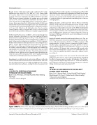
Methods: In lambs, a pulmonary valve stenosis (PS) was created by placing a PTFE strip around the pulmonary artery through a right thoracotomy (Figure A). Four months later a bare metal stent was anchored across the pulmonary valve in the PTFE strip (Figure B) thereby relieving the stenosis and creating pulmonary valve insuf- ciency (Figure C). Melody valve implantations into this bare metal stent at di erent time intervals are ongoing (Figure D). Follow up by means of MRI was performed to assess remodeling and reversed remodeling of the RV.
Results: All animals survived the initial surgical phase (n=9). Two ani- mals died during bare metal stent implantation (ventricular brilla- tion n=1; PA rupture by balloon dilation n=1). MRI showed signs of RV hypertrophy prior to relief of stenosis compared to healthy controls. Total RV cardiac output (CO) was 2.2±0.7 L/min after PS, 5.0 ±0.8 L/ min immediately after bare stent implantation and 3.5±0.1 L/min SD after 5 months of PR. Animals had an important PR 5 months after bare metal stent implantation (32±2.3%). The LV-RV EDV ratio was 0.7±0.1. This was signi cantly smaller compared to healthy controls (p<0.05)(n=3).
Conclusion: The creation of an ovine survival TF model with initial pulmonary valve stenosis and secondary regurgitation (mimicking the e ect of surgical repair) is feasible. All hallmarks of the TF phys- iology (ventricular hypertrophy after stenosis – ventricular dilatation due to PR) were realized. This model forms the ideal basis to study the timing of pulmonary valve replacement in TF.
#0078
10 YEARS OF EXPERIENCE WITH THE MELODYTM VALVED STENT FOR PPVR
Bjorn Cools1, Werner Budts1, Derize Bosho 1, Ruth Heying1, Ward Vanagt2, Els Troost1, Stefan Frerich2, Marc Gewillig1 1UZ Leuven, Leuven, Belgium
2AZ Maastricht, Maastricht, The Netherlands
AFi/gPuTrFeE1s(t#r0ip0s7u7p).raP-aannenlAu.laPrTFaEnsdtrmipasrukperai-nanfunnudlaibraunladrmB/aMrkeRrIisnhfuonwdiinbgulbaar.rPeamneelBta.MlsRteIsnhtoawcicnrgosbsatrheemneata lvsetentacrossthenative TM
pulmonarryvvaalvlvee.PCa/nMelRCI.MphRaIspehsahseirsthairxtisaxsihsoswhoinwginmgamssaisvseivPeRPRC./PdaenpelloDy.eDmepelnotyeomfMenetlofdtyheMvealovdeyiTMnvbalrvesintebnatrestent. Journal of Structural Heart Disease, December 2016 Volume 2, Issue 6:241-306
