
Page 76 - Journal of Structural Heart Disease Volume 2, Issue 6
P. 76
303
Meeting Abstracts
Joseph DeGiovanni1, C Mehta2, P Noonan3, P Clift1, V Grech4, I Spadoni5
1University Hospital Birmingham, Birmingham, UK 2Birmingham Children’s Hospital, Birmingham, UK
3Yorkhill Children’s Hospital, Glasgow, UK 4Mater Dei Hospital, Malta, Malta
5Heart Hospital, Massa, Italy
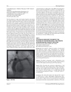
The nal palliation for single ventricle physiology these days utilises the surgical technique of Total Cavo Pulmonary Connection (TCPC) using a ptfe conduit between the inferior naval vein and the pulmo- nary arteries. This concept is considered to favour haemodynanics and reduce atrial arrhythmias. Whilst surgical fenestrations are some- times performed these can get occluded; in those where there is no fenestration, entry into the LA mass through the conduit is not easy, sometimes impossible and not without risk. We describe a technique to enter the atria mass (AM) from the right internal jugular vein, mainly using a transeptal needle. The proximity of the pulmonary artery (PA) just to the left of the conduit anastomosis and the left atrium makes this feasible, it means crossing native tissue only and previous sur- gery and adhesions make the procedure safe. The procedure is car- ried out under general anaesthesia and with TEE guidance. a CT prior to the procedure helps with a better understanding of the anatomy although this is not mandatory. Angiography is carried out simulta- neously from the superior naval vein and the left atrium (entered ret- rogradely via the aorta and ventricle through the AV valve). This helps with the puncture site, direction of needle and distance between the PA and AM. Pressure monitoring is important during puncture. Once the AM is entered and con rmed on pressure, TEE and angiography, the planned procedure can proceed after heparinisation. We carried out this procedure in 6 patients, 5 children and 1 adult. The ages of the children ranged from 4 to 11 years and the adult was 30 years old. In 4 of the cases, the procedure was mainly carried out to create
Figure 1 (#0150).
a stent fenestration, 2 within weeks of the TCPC and 2 much later for protein losing enteropathy (PLE). In 2 patients, concomitant IVC/con- duit anastomosis stenting was carried out one with a CP stent and one with an Optimus stent because of documented stenosis. One older child required access to the AM for electrophysiology and radio frequency ablation and the adult patient required a permanent trans- venous atrial pacing lead because of exit block on a chronic atrial lead which was problematic to implant many years earlier. The pro- cedures were all successful. In one patient, there was early closure of the stent fenestration and this was initially managed with i.v. TPA and once ow was reinstated, the stent was dilated with a ballon and has remained open an with no recurrence of the PLE. The stent used was an Andrastent XXL as this is what was available but these stents are designed for larger diameters. An “X” model would have been better. Access to the AM in TCPC Fontan is increasing, in part as this popula- tion is rising, they are living longer and hence will develop problems with age. Although we have used this technique safely and showed proof of concept for fenestration, permanent transvenous pacing and for arrhythmia therapy, other indications will arise in future, such as AV valve repair, left atrial appendage occlusion, pulmonary vein isola- tion and paravalvular leaks. Potentially, we could also see indications for percutaneous or hybrid AV valve replacement which will make this technique essential in TCPC physiology.
#0151
UTILIZING FLUOROSCOPY TO IDENTIFY THE POSITION OF THE ENDOTRACHEAL TUBE PRIOR TO CATHETERIZATION: A QUALITY INITIATIVE
Daniel Gruenstein, Melissa Webb, Ala Soo an,
Shari Slyder, Caitlyn Aveyard
Daniel Gruenstein, Chicago, IL, USA
Background: Unrecognized or delayed recognition of endotracheal tube malposition during cardiac catheterizations for congenital heart disease may lead to avoidable complications in the cath lab. Consequences include inaccurate hemodynamic data from hypoven- tilation, or patient complications from inadequate gas exchange. A quality initiative of uoroscopically verifying and correcting endo- tracheal tube (ETT) position before obtaining hemodynamics during catheterization was implemented. We describe the frequency of ETT repositioning with this practice.
Methods: All patients undergoing cardiac catheterization since implementing this practice were retrospectively reviewed. Primary outcome of the study was the frequency of ETT repositioning.
Results: Of the 66 patients who underwent catheterization, 3 had a tracheostomy tube or ETT already in place. Of the remaining 63 patients, 17 (27%) required repositioning due to high or low place- ment. The mean weight and age for those requiring repositioning was 15.0 kg (2.9 – 78) and 3.3 years (0.1 – 20) and for those who did not require repositioning was 23.3 kg (3.1 – 104.8) and 5.7 years (0 – 37) respectively.
Conclusions: Our study demonstrates that improper ETT position is common prior to cardiac catheterization. Implementing a uo- roscopic check and correction of the ETT position prior to starting the procedure is simple, low risk, and it may improve the quality of hemodynamic data and avoid preventable complications. If broader
Hijazi, Z
20th Annual PICS/AICS Meeting Abstracts
