
Page 10 - Journal of Structural Heart Disease Volume 3, Issue 2
P. 10
Original Scienti c Article
36
Materials and Methods
Study Design
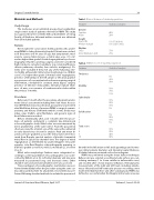
Table 1. Clinical features of studied population.
Features
Statistical analysis
The study was an uncontrolled, prospective, longitudinal, single-center study of patients referred for PBMV. This study was approved by the Scienti c Ethical Committee of Sohag Faculty of Medicine. Informed written consent was obtained from all included patients.
Gender
Females Males
Height
Mean (SD) Median (range)
Rhythm
Sinus
Atrial brillation (AF)
80% 20%
159.57 (8.50%) 159 (145-178%)
75% 25%
Patients
We recruited 40 consecutive elderly patients who under- went PBMV in Sohag University Hospital. Patients were includ- ed if they were over 60 years of age; had symptomatic mod- erate to severe mitral stenosis, a mitral valve area <1.5 cm2, and no higher than grade 2/4 mitral regurgitation by echocar- diography; refused to undergo surgery; and were considered high risk due to comorbid conditions (e.g., renal insu ciency, chronic pulmonary disorder, liver cirrhosis, malignancy). Pa- tients were excluded if they met any of the following crite- ria: highly unfavorable mitral valve morphology (i.e., Wilkin’s score ≥11), higher than grade 2/4 mitral valve regurgitation, presence of thrombus in the left atrium or left atrial append- age, presence of concomitant valve disease requiring surgical intervention, scheduled for coronary artery bypass surgery, presence of infective endocarditis, interatrial septum thick- ness >4 mm, or occurrence of cerebrovascular stroke within the previous 3 months.
Procedures
Before and 1 month after the procedure, all patients under- went clinical assessment including New York Heart Associa- tion (NYHA) functional classi cation, paroxysmal or persistent atrial brillation, history of previous PBMV or surgical commis- urotomy, and history of thromboembolic events. Body mass index, sinus rhythm, atrial brillation, and general signs of heart failure were recorded.
Before, immediately after, and 1 month after the proce- dure, all patients underwent a complete two-dimensional echocardiographic study. Mitral valve area was measured by direct plannimetry of the valve ori ce. From the parasternal short-axis view, the smallest area of the valve ori ce obtained in early diastole was chosen for analysis. Peak and mean di- astolic pressure gradients across the mitral valve were mea- sured from Doppler spectral analysis of diastolic transmitral mitral ow in apical four-chamber view. The degree of mitral regurgitation as determined by pulsed Doppler echocardi- ography, color- ow Doppler echocardiography mapping, or both was graded as none (0), mild (1), moderate (2), or severe (3 or 4).
Mitral valve morphologic features were categorized ac- cording to a semi-quantitative echocardiographic score as de- scribed by Wilkin’s et al. [6]. Left atrial diameter and size were measured in parasternal long- and short-axis views. Other chamber sizes and valve abnormalities were also assessed.
Transesophageal echocardiographic examination was per- formed immediately before the procedure using the same car- diac ultrasound machine for all patients. Assessment was per- formed following standard methods to exclude the presence of
Table 2. Willkin’s Score of studied population.
Thickness
1 5% 2 45% 3 50%
Mobility
1 10% 2 75% 3 15%
Subvalvular
1 10% 2 50% 3 35% 4 5%
Calci cation
1 5% 2 50% 3 35% 4 5%
Total score
7 10% 8 20% 9 40% 10 30%
thrombi in the left atrium or left atrial appendage and to mea- sure mitral annular diameter and interatrial septal thickness. A blinded observer reviewed each echocardiogram.
PBMV was performed using the Inoue balloon technique. Balloon size was selected according to body surface area con- sidering anatomy (1 to 2 mm smaller in unfavorable cases) and reached after several stepwise in ations. Hemodynamic measurements of the right and left heart, including simulta- neous left atrial and left ventricular pressure recordings, were made immediately before and after valvuloplasty. PBMV was de ned as successful when the Doppler mitral valve area was
Features
Statistical analysis
Journal of Structural Heart Disease, April 2017
Volume 3, Issue 2:35-42
