
Page 14 - Journal of Structural Heart Disease Volume 3, Issue 3
P. 14
67
Review Article
cedures allows the stent to embed and to get a better idea of diameter in the event of recoil. If the latter is dynamic or signi cant (arbitrarily, a gradient of great- er than 20 mm Hg), a second stent may be required. Right ventricular out ow tract preparation must be done prior to valve implantation and do not rely on post dilatation after the valve is implanted.
Procedure
Pulmonary valve implantation can be carried out under sedation although most are carried out under general anesthesia. A biplane system is preferable and the room must have adequate levels of sterility and air change to minimize risk of infection. The operators must adhere to strict sterile protocols throughout. Antibiotics are generally given to cover the proce- dure mainly to cover staphylococcal infections but this is not a substitute to good operator and patient preparation. The procedure is covered with heparin in therapeutic doses based on ACT measurement.
The femoral vein is accessed percutaneously high up to enter the external iliac vein. If the femoral veins are occluded or tiny or if there is absence of the IVC with azygos replacement, a right internal jugular ap- proach is required. The jugular approach may also be preferred in patients with TGA or ccTGA.
A 5–6 mm skin incision is made at the entry site and if a closing device is planned this should be in- serted at this stage. It is customary to place a 16–18 Fr short sheath to allow for angiography, balloon in- terrogation, or stenting of the RVOT if required. Right sided pressures are recorded and a right ventricu- logram carried out to show the RVOT anatomy and pulmonary arteries, as this helps to determine which pulmonary artery to park the exchange wire in or- der to optimize the catheter course. In general, the left pulmonary artery is preferred when approaching from the femoral and the right pulmonary when ap- proaching from the jugular but there are no hard and fast rules as this depends on the anatomy, approach, and operator preference. Arterial access with a 4–6 Fr sheath is obtained.
Once the RVOT is prepared with stenting, the PPVI procedure can proceed. Having a sti ex- change-length guide wire in a peripheral pulmonary artery is essential and this is placed through a MP
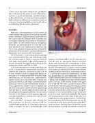
Figure 3: Animation of pulmonary valve implantation with the Edwards valve.
catheter; a Lunderquist 0.035” wire is commonly used. Over the wire, an appropriate Edwards introducer sheath, often nowadays this is the eSheath, is insert- ed having pre-dilated the track with the Edwards di- lator kit. The Edwards XT or Sapien 3 valve is loaded and crimped on to the NovaFlex/Commander delivery system proximal to the balloon and making sure that it is orientated for pulmonary implantation—at least two people must see and con rm this. Choose the correct crimper for the valve size. The delivery system is then passed through the introducer sheath until the valve is well out of the delivery sheath in the upper part of the IVC. The valve is pushed forward on the proximal part of the balloon by releasing two catches on the delivery handle and pushing the ared sheath forward monitoring this on uoroscopy - during this maneuver, it is essential to keep an eye on the wire position to avoid inadvertent withdrawal. Fine tuning of the valve position on the center of the balloon as judged by the radio-opaque markers, is achieved by the rotating knob on the delivery handle. It is best for this maneuver to be done in the IVC and not the RVOT. Once the valve is optimally placed on the balloon, the whole assembly is pushed over the wire towards the
DeGiovanni, J.
Pulmonary Valve Replacement
