
Page 21 - Journal of Structural Heart Disease Volume 3, Issue 3
P. 21
Meeting Abstracts
74
24 patients (82.7%). Implantation failed in ve patients due to de - cient aortic rim in two patients and increased severity of AR in the other three patients.
Conclusion: Transcatheter closure of PM VSD with SAR using ADO type I is safe and e ective with the very low incidence of complications.
STAGED SWISS CHEESE VSD CLOSURE
Mylah Alfeche
University of the Philippines-Philippine General Hospital, Manila Philippines
An 8-year old male with cleft lip and palate presented with multiple muscular ventricular septal defects (VSD) diagnosed since 6 months of age. Anti-heart failure medical treatment with furosemide and digoxin were commenced by another cardiac center and was con- tinued until the present time. Financial constraints prevented the performance of surgical correction, which was advised on numerous occasions.
2D echocardiogram showed multiple muscular VSDs with the most prominent being two muscular VSDs, measuring 7-8 mm and 5-6 mm located near the apex. There was some left to right shunting, left heart enlargement and signs of severe pulmonary hypertension (severe tricuspid regurgitation, right ventricular hypertrophy). He was planned for transcatheter closure of these 2 larger defects.
Under general anaesthesia and 100% oxygen support, the pulmo- nary artery pressure was documented at (103/50 mmHg, mean of 75 mmHg) 80% of systemic pressure. The Qp/Qs was 3:1 on oxygen. Left ventricular angiograms showed a midmuscular VSD (LV side 9.4 mm, RV side 6.6 mm, length 11.9 mm) and apical VSD (6.6 mm). The midmuscular VSD was accessed using a modi ed pigtail catheter. An AV loop was created from the femoral vein and the VSD was closed in standard fashion using a Cocoon VSD 12-7 mm through an 8F sheath. The position of the device was con rmed by angiography and trans- thoracic echocardiography. The apical VSD was crossed antegradely from the RV accessed from the internal jugular vein using a JR4 cath- eter over a glidewire with the tip positioned in the descending aorta. A 7F ductal delivery sheath was advanced from the right IJ over the wire and a 10-7 mm Cocoon VSD occluder was deployed under uo- roscopic and echocardiographic guidance. The patient tolerated the procedure without untoward events and was discharged with the fol- lowing medications: aspirin (4 mg/kg/day), sildena l (2 mg/kg/day), furosemide (0.5 mg/kg/day), captopril (1 mg/kg/day) and digoxin (10 mcg/kg/day). Repeat echocardiogram two days after the procedure showed the two devices in place without leak, but there were still another two muscular VSDs (4.5 mm and 6 mm) located between the two Cocoon VSD devices.
After two years from the transcatheter closure of the 2 muscular VSD, he was readmitted for transcatheter closure of the remaining muscu- lar VSDs. Hemodynamic study on 100% oxygen showed the PA pres- sure at 90% systemic. The Qp/Qs was 2.2:1 with a PVR of 3.9 Wood units/m2. Left ventriculogram revealed two midmuscular VSDs mea- suring 5.8 mm (superior) and 5.5 mm (inferior).
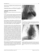
Figure 1.
The two VSDs were crossed and closed sequentially in standard fash- ion using a 6-7 mm Cocoon VSD occluder in each VSD. There was still some small residual VSD after these residual VSDs were closed, as is expected in this type of VSD.
Figure 2.
The child tolerated the procedure without untoward events and was discharged with the following medications: aspirin, sildena l, furo- semide, captopril and digoxin. This case demonstrates the feasibility and safety of staged closure of Swiss cheese VSD using the Coccon device.
Journal of Structural Heart Disease, June 2017
Volume 3, Issue 3:73-95
