
Page 25 - Journal of Structural Heart Disease Volume 3, Issue 4
P. 25
Case Report
112
the IVC course.
The procedure was performed under general
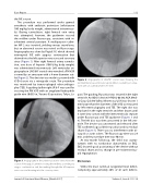
anesthesia with antibiotic protection (cefotiaxone 100 mg/kg body weight, administered intravenous- ly). During cannulation, right femoral vein entry was attempted; however, the guidewire crossed the midline under uoroscopy, consistent with in- advertent arterial puncture. A multipurpose cathe- ter MP 2 was inserted, yielding venous waveforms, but an abnormal course was noted on uoroscopy. Angiography was done by Pigtail 5-F, which showed interrupted IVC with azygous continuation that drained into the left superior vena cava and coronary sinus (Figure 1). After right femoral artery cannula- tion, one dose of heparin (100 U/kg body weight) was administered intravenously. Left ventricular an- giography in LAO/60° cranial view revealed a PM VSD covered by an aneurysm with a 4-mm diameter out- let (Figure 2). The decision was made to proceed with VSD closure via a retrograde route. The procedure was monitored by transesophageal echocardiogra- phy (TEE). A guiding Judkin right JR 6-F was used for crossing the PM VSD with an angulated hydrophilic guide wire (0.035-in, Terumo Corporation, Tokyo, Ja-
Figure 1. Angiography in the anterior-posterior view shows the course of the pigtail catheter crossing the midline, ascending via azygos continuation, and descending through the left superior vena cava and draining into the coronary sinus.
Figure 2. Angiography in LAO/60° cranial view showing the perimemberanous ventricular septal defect covered by an aneu- rysm with an outlet diameter of 5 mm.
pan). The guiding JR position was secured in the right ventricle. An ADO II device (9-PDA2-06-04, AGA Medi- ca Corp, Golden Valley, Minnesota, USA) was chosen 1 mm larger than the diameter of the VSD as measured by left ventricolography and TEE. The right disc was deployed in the right ventricle and then drawn until it came into contact with the interventricular septum under uoroscopy and TEE guidance (Figures 3 and 4). The left disc was then uncovered in the left ven- tricle. The device was unscrewed and released after TEE con rmed a good device position and no residual shunt (Figure 5). There was no interference with tri- cuspid or aortic valves. The uoroscopy time was 24 min, and the procedure time was 90 min.
At two-month follow-up, the child was asymp- tomatic with no conduction abnormality on ECG. ECG showed good positioning of the device without residual shunt and no change in pre-existent tricus- pid regurgitation.
Discussion
VSD is the most common congenital heart defect, comprising approximately 20% of all such defects.
Journal of Structural Heart Disease, [Month 2017]
Volume 3, Issue 4:111-114
