
Page 21 - Journal of Structural Heart Disease Volume 3, Issue 6
P. 21
177 Case Report
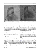
Figure 1. Panel A. Cardiac angiography prior to stent placement. The con uence narrowing is 3.6mm with the superior aspect of the vertical vein measuring 5.5mm. Panel B. Cardiac angiography after the two stents were implanted, with the second stent telescoped proximally within the initial stent.
cyanosis, and metabolic acidosis [4]. It is traditionally considered a surgical emergency. Emergent stenting of the VV is rarely reported but has been performed when a patient is considered a poor candidate for surgical repair [4-6]. Pulmonary venous obstruction is associated with poor prognosis and a high risk of operative mortality [7, 8].
Here, we report a case of an infant patient who was electively referred to catheterization for stent place- ment to relieve VV stenosis. Our objective was to prevent the emergent need for surgical intervention while allowing additional growth before surgery.
Case Presentation
A 7-day-old, former 36 6/7 week, 2.4 kg, small for gestational age male infant was transferred from an outside institution. At the time of delivery, he was not- ed to have poor respiratory e ort and a heart rate be- low 100 beats per minute. Chest compressions were provided for less than 1 min, and his APGAR scores were 5 and 7 at 1 and 5 min, respectively. The patient was noted to have oxygen saturation of roughly 80%, requiring blow by oxygen. He was initially placed on
nasal cannula, but over the next 7 days progressed to continuous positive airway pressure and had a chest X-ray suggestive of pulmonary edema. An echocar- diogram at that time revealed supracardiac TAPVC, a small apical muscular ventricular septal defect with right-to-left shunt, and a moderate secundum atrial septal defect with right-to-left shunt.
On admission, the patient was stable with a base- line oxygen saturation of 72% on 40% oxygen. An echocardiogram was performed that con rmed su- pracardiac TAPVC but demonstrated an obstruction in the VV with a mean gradient of 22 mmHg. All pul- monary veins drained to a con uence behind the left atrium and communicated to the innominate vein via aVV.
The patient’s size and the desire to optimize his clinical stability led to the decision to undergo car- diac catheterization for stent implantation in the VV (Figure 1A). Femoral vein access was achieved, and a 4-F angled Glide catheter (Terumo, Somerset, NJ, USA) was advanced prograde into the VV. A 17 mmHg pres- sure gradient between the pulmonary venous con- uence and the left innominate vein was recorded. A V-18 Control wire (Boston Scienti c, Marlborough,
Rhee E. et al.
Elective Stent Implantation for TAPVC
