
Page 13 - Journal of Structural Heart Disease Volume 4, Issue 1
P. 13
Original Scienti c Article 4
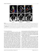
Figure 3. Series of color (Panels A, B, C) and two-dimensional transthoracic echocardiography four-chamber view images performed at baseline (Panels A, D), 1-month follow-up (Panels B, E), and 6-month follow-up (Panels C, F) in a 20-year-old female patient after surgi- cal closure of ventricular septal defect and right ventricle out ow obstruction using a patch. She had severe pulmonary regurgitation. These images demonstrate sustained resolution of free pulmonary regurgitation and signi cant remodeling of the right ventricle. The white arrow in the Panel A image indicates severe pulmonary regurgitation, and the images in Panels B and C demonstrate a competent Venus P-valve (26 mm) with trivial if any regurgitation. The Panel D image shows a large right ventricle, and the images in Panels E and F show signi cant remodeling of the right ventricle at 1- and 6-month follow-up, respectively.
Procedure and Follow-Up
It is advisable to perform the procedures under general endotracheal anesthesia. Access should be via both right and left femoral veins (right femoral for device deployment and left for angiographic control), and the femoral artery should also be accessed to as- sess coronary artery proximity to the RVOT. Heparin 100 U/kg should be administered to maintain acti- vated clotting time of >250 s. Intravenous antibiotics should be given at the beginning of the procedure followed by two doses 8 hours apart. Transesopha- geal echocardiography (TEE) or intracardiac echocar- diography can be performed during the procedure to monitor the RVOT before and after valve implan- tation. However, this step is optional. Hemodynamic assessment and detailed angiography including bal- loon sizing can be repeated if needed. A 260-cm long 0.035” Lunderquist extra-sti guide wire (Cook Medi- cal, Bloomington, Indiana, USA) or any other sti wire
can be positioned in the PA, preferably in the distal left lower PA branch. Intended valve diameter selec- tion should be 2–4 mm larger than the balloon infla- tion diameter at its waist, whereas mid-body length selection should be equivalent to the distance from the RVOT to PA bifurcation.
After preparation of the valve by rinsing with 2,000 ml normal saline for at least 10 min, the valve is man- ually crimped in a bath of cold normal saline onto a 20–22-F delivery system. The valve assembly is then passed through a 22-F Check-Flo® Performer Ex- tra-Large Introducer sheath (Cook Medical) and ma- nipulated over the Lunderquist guide wire. The distal carrot tip of the assembly then is advanced into the proximal left PA. Frequent check angiograms through a pigtail catheter placed in the main PA should be done as the distal flare of the valve is slowly deployed by clockwise rotation of the releasing knob. The valve position can be adjusted after check angiograms
Journal of Structural Heart Disease, February 2018
Volume 4, Issue 1:1-8
