
Page 26 - Journal of Structural Heart Disease Volume 4, Issue 3
P. 26
83 Original Scienti c Article
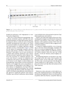
Figure 3. Rate of change in Pulmonary Artery Aneurysm size in 11 patients over time. The bold and dashed line is the mean rate of growth in the entire cohort over 10 years.
progressive pulmonary valve regurgitation or coro- nary artery compression.
Niwa et al. [9] described the histopathologic char- acteristics of thirteen patients with PAA and com- pared the ndings to seventy-three patients with aor- tic aneurysms due to a variety of causes. One patient with PAA and pulmonary valve stenosis was includ- ed and had evidence of advanced (grade 3) medial wall abnormalities of collagen deposition, ground substance and loss of elastin bers (Figure 1). There are rare reports of aortopathy in patients with pul- monary valve stenosis and PAA. In 1959, Evans and Dauncey [7] described a case of aortic dissection in a patient with moderate pulmonary valve stenosis with “post-stenotic” pulmonary artery dilation. How- ever, the occurrence of congenital pulmonic and aortic valve disease is unusual. In congenital aortic or pulmonic valve stenosis, the relationship of sever- ity of stenosis to the degree of arterial dilation and histopathologic abnormalities has been questioned. Niwa et al. [9] suggest that aortic and pulmonic ar- terial dilation are independent of the degree of val- vular stenosis or regurgitation and more likely due to an inherent congenital arteriopathy. This assertion is supported by the eleven patients described in this cohort, all of whom had pulmonary artery dilation in the absence of signi cant pulmonic valve stenosis. Therefore, “post-stenotic” pulmonary arterial dilation
is not included in this cohort given the absence of sig- ni cant pulmonary valve stenosis.
Patients were generally diagnosed because echo- cardiographic imaging was performed to evaluate for pulmonic valve ejection sound and/or systolic ow murmurs in the pulmonic position. There were two incidental diagnoses made on chest imaging to eval- uate other conditions.
In regard to imaging modalities, it was encourag- ing to note that echocardiography correlated well with cross-sectional imaging by MRI or CT. Echocar- diography is widely available, cost-e ective and does not involve radiation. Given the slow rate of PAA growth (~ 5 mm over 10 years), yearly imaging is un- necessary.
The following limitations are present in our study. The sample size for this study is eleven patients. No de nitive claims can be made with a small sample size. This study is also retrospective. Even though the current progression of the patients in our study is good, we can’t be sure that they will continue on the same trajectory.
Conclusions
Pulmonary artery aneurysms are described in pa- tients with congenital pulmonary valve pathology of mild functional signi cance. Most cases were present in patients older than 50 years. Long-term follow-up
Obeid K. et al.
Pulmonary Arteriopathy without Valvular Anomalies
