
Page 42 - Journal of Structural Heart Disease Volume 4, Issue 3
P. 42
99
Meeting Abstracts
Intervention:
Case 1: After hemodynamic assessment, femoral arterial access was replaced with a 14-Fr sheath in a surgical “cut- down” manner, and a stent (P4010) was placed to cover the proximal end of the rst stent. Because the additional stent remained incompletely apposed to both the anterior aortic wall and subclavian artery, a Coda 32-mm balloon (Cook Medical, Bloomington, IN) was advanced to the proximal end of the second stent and manually in ated to prevent stent mal-apposition and partial jailing of the left subclavian artery. Angiography and hemodynamic evaluation after the post-dilatation using the Coda balloon demonstrated complete apposition to the vessel wall and no measurable residual gradient.
Case 2: After placing a 14-Fr sheath at femoral vein, post-dilatation using a Coda balloon 32-mm was per- formed for the proximal end of the stent. Angiography after the procedure demonstrated complete apposition to the anastomosis. Both procedures were uncomplicated. The stent was brie y and successfully apposed to vessel wall with origin of side branch vessels opened, relieving obstruction and deformity of the vessel.
Learning Point of the Procedures: Aortic coarctation (CoA) and pulmonary stenosis (PS) are often located in curved segment and adjacent to the origin of other vessels. Stenting for such lesions is sometimes accompanied by stent mal-apposition and partial jailing of side branch ves- sel, which may be related with thrombosis or branch vessel occlusion. We performed post-dilation using Coda balloon to appose the stent to vessel wall. Coda balloon is a spher- ical semi-compliant balloon catheter intended for tempo- rary occlusion of large vessel and post-dilatation of stent graft in adult. Apposition using Coda balloon is a quite simple and e ective resolution in stenting for CoA and PS.
19. IMPERFECT PDA STENTING IN A BABY GIRL WITH TETRALOGY OF FALLOT
Ming-Tai Lin1, Hsin-Chia Lin2, Jou-Kou Wang3
1 Department of Pediatrics, National Taiwan University Children Hospital, Taiwan; Pediatric Interventional Cardiology, Kawasaki Disease ; Pediatric Cardiology
2 National Taiwan University Hospital; Pediatrics; Cardiology
3 National Taiwan University Children Hospital; Interventional Cardiology; Pediatric Cardiology
History and Physical: 7-day-old female baby was a victim of tetralogy of Fallot with small patent ductus arteriosus (PDA). After delivery, cyanosis (SpO2=75-80%) was noted with a Gr II/VI systolic murmur at her left middle sternal border. Prostaglandin E1 (PGE1, 5 ng/kg/minute) was given to maintain adequate pulmonary blood ow.
Imaging and Indication: Computed tomography demon- strated small pulmonary arteries (McGoon index:1.0). Therefore, we plan to do PDA stenting for her rst-stage operation.
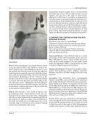
Intervention: Left innominate artery cineangiogram showed a small PDA connected between left subclavian artery (LSCA) and bi cation of bilateral pulmonary arteries. (Figure A) We passed a Rinato coronary wire to his distal LPA via the PDA and then deployed a REBEL (4 x 8 mm) and a MULTI-LINK8 (3.5 x 15 mm) stent at his PDA. However, com- promised LSCA ow and thrombosed stent (no ow) were noted 10 minutes after stent implantation. NC QUANTUM APEX balloon (4 x 15 mm) was advanced to redilate the two stents and successfully restore the ow.
Another Rinato wire was advanced to left innominate artery, through the mesh of the implanted stents, nally to her LSCA. NC Sprinter balloon (3.5 x 15 mm) was used to dilate the mesh at the origin of LSCA. Cineangiogram con rmed the patency of LSCA origin. (Figure B) She was
Hijazi, Z
2017 CSI Africa Abstracts
