
Page 55 - Journal of Structural Heart Disease Volume 4, Issue 3
P. 55
Meeting Abstracts
112
signi cant PDA and received pharmacological therapy as protocols. Median age and weight for procedure was 22 days (2-133 days) and 1,500 g (478-2,350 g), respectively. Six patients were associated massive pulmonary haemor- rhage. The mean minimal ductal diameter was 3.5 + 1.1 mm. The mean ductal length was 7.8 + 3.6 mm. The most common PDA type was type C (n=7). There were 3 times of device migration before detachment which required to retrieve and deploy a new one. Devices used in this study were AMPLATZERTM duct occluder II additional size (ADO II AS) (n=13), AMPLATZERTM vascular plug I (VP 1) (n=1), and vascular plug II (VP 2)(n=3). Complete closure were achieved in all patients except one patient had progres- sive coarctation which required surgical removal 4 months later.
Conclusions: It is now currently feasible to undertake tran- scatheter PDA closure in premature infants body weight greater than 478 grams according to our experiences. We added at least 1.5 mm in minimal ductal diameter by echo- cardiogram to select the device waist. We concluded that ADO II AS could be best used in ductal diameter less than 3.5 mm, and VP 2 could be used in larger and long ductus, while VP 1 used in large and short (less than 10 mm) one.
37. TRANSCATHETER CLOSURE OF DOUBLE VENTRICULAR RUPTURE IN A PATIENT WITH NON-ST ELEVATION MYOCARDIAL INFARCTION
Tse-Hsuan Yang
Kaohsiung Veterans General Hospital, Taiwan; Cardiovascular Center; Cardiology
History and Physical: A 68-year-old lady has history of hyper- tension, type 2 diabetes mellitus and uremia with regular hemodialysis, presented with sudden acute of chest pain on Jun. 1st 2015. Non-ST-elevation myocardial infarction was diagnosed, and percutaneous coronary intervention (PCI) with drug-eluting stent was performed over the prox- imal portion of left anterior descending (LAD) artery. Three days after PCI, apical ventricular septal rupture (VSR) was diagnosed because of grade 3 holosystolic murmur at the apical area. Three weeks after PCI, surgical repair for VSR was performed smoothly and she was discharged asymp- tomatically. Three months after surgical repair, she experi- ence exertional dyspnea and e ort-related chest pain. The CT angiography showed a left ventricular pseudoaneu- rysm over the middle portion of anteroseptum with con- nections to RV. The coronary angiography showed di use atherosclerosis over distal portion of left anterior descend- ing artery with dynamic compression of LAD by the pseu- doaneurysm. After thorough evaluation of the heart team,
percutaneous coronary intervention, transcatheter closure of VSR and pseudoaneurysm were proceeded.
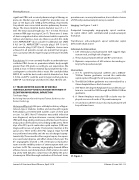
Imaging: See Figures 1 and 2.
Computed tomography angiography: apical ventricu- lar septal defect with communicated pseudoaneurysm fromation
Transthoracic echocardiogram: apical ventricular septal defect with shunt noted
Indication for Intervention:
1. Left ventricular pseudoaneurysm with ragged edge,
narrow neck, and high risk of rupture
2. The pseudoaneurysm with LV and RV connections
3. Dynamic compression of left anterior descending ar- tery by the pseudoaneurym
Intervention:
a. A 5 Fr. cutted-head pig-tail catheter and a 0.032in-
*260cm Terumo guidewire crossed the ventricular septal rupture through the LV pseudoaneurysm.
b. The 0.032in*260cm guidewire was externalized by a 25mm Amplatz Goose Neck Snaire kit.
c. A 8F 80cm (45 degree) Amplatzer Torvue delivery sys- tem was crossed the VSR through the 0.032in*260cm guidewire.
d. A 16mm Amplatzer muscular VSD occluder was de- ployed over one of necks of LV pseudoaneurysm
e. Second transcatheter closure of pseudoaneurysm will be performed soon.
37. Figure 1.
Journal of Structural Heart Disease, April 2018
Volume 4, Issue 2:85-113
