
Page 19 - Journal of Structural Heart Disease Volume 5, Issue 1
P. 19
Case Report
8
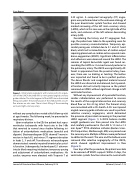
Figure 1. Initial lateral angiogram, with cranial and LAO angula- tion of the LMCA obtained with a non-tapered angled coronary guide catheter. The initial segment of the LMCA has a diminished filling and caliber. The proximal part of the LAD and the circum- flex arteries are also seen. There is back filling of the ascending aorta and right coronary artery.
without any complications noted at his follow-up visit at age 8 weeks. The following week, he presented in respiratory distress.
Upon evaluation in the ED, the patient had supra- ventricular tachycardia with heart rate in the 160s that reverted to normal sinus rhythm following ini- tiation of antiarrhythmic medications (esmolol and digoxin). Electrocardiogram (ECG) showed T-wave in- version in lead aVL and minor ST segment elevation in leads 2, 3 and aVF. Transthoracic echocardiogram demonstrated severely impaired biventricular systol- ic function. Subsequently, he developed 2:1 atrioven- tricular block requiring intubation, temporary pacing and adjustment of antiarrhythmic medications. His cardiac enzymes were elevated with Troponin T at
2.23 ng/mL. A computed tomography (CT) angio- gram was performed due to the unknown etiology of the poor biventricular systolic function and showed marked narrowing of the left main coronary artery (LMCA), which in this case was on the right side of the aorta, and occlusion of the left anterior descending artery (LAD).
Considering the history and CT angiogram find- ings, the patient was taken to the operating room for possible coronary revascularization. Temporary epi- cardial pacing was initiated due to 3:1 and 4:1 heart block, which led to immediate loss of cardiac output requiring placement on cardiac extra corporeal mem- brane oxygenation (ECMO). Significant inflammation and adhesions were observed around the LMCA. The remains of topical hemostatic agent was found sur- rounding the LMCA as it curved around posterior to the pulmonary artery. The LMCA was significantly dif- ferent in appearance from the proximal artery, how- ever, there was no kinking or twisting. The button was inspected and found to be in perfect position. The dense fibrotic and coagulated material around the LMCA was dissected and released, but myocardi- al function did not instantly improve so the patient remained on ECMO without significant change in left ventricular function.
Without any improvement of myocardial function, cardiac catheterization was performed to reassess the results of the surgical intervention and coronary blood flow on this 4.5 kg infant. The femoral artery was cannulated with a 4 Fr sheath. A 4 Fr non-tapered angled coronary guide catheter was used to perform selective coronary angiograms, which delineated the presence of persistent narrowing in the proximal LMCA segment (Figure 1). A 0.014 balance middle weight guidewire was then passed into the LMCA and carefully positioned in the LAD. A 1.5 mm x 12 mm ApexTM PTCA dilatation catheter (Boston Scien- tific Corporation, Marlborough, MA) was passed over the coronary wire. Multiple inflations were performed to obliterate the waist. The wire and the balloon were removed after selective angiography of the LMCA, which showed significant improvement in flow (Figure 2).
Four days after the procedure, the patient was able to be weaned off ECMO support. Within 9 days, the ventricular systolic function returned to normal with
Journal of Structural Heart Disease, February 2019
Volume 5, Issue 1:7-10
