
Page 23 - Journal of Structural Heart Disease Volume 5, Issue 1
P. 23
Case Report 12
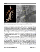
Figure 1. Panel A. Pre-procedure computed tomography scan and Panel B. Pre-procedure angiogram showing the Glenn anastomo- sis with superior vena cava connected to the right pulmonary artery (A=14 mm); Fontan conduit (inferior vena cava, B=18 mm; left pulmonary artery C=13 mm). The distance between the disconnected Glenn and Fontan anastomoses is D=5 mm.
Gortex aorto-pulmonary shunt with narrowing of the Norwood anastomosis. She then underwent surgical revision of the Norwood anastomosis. A classic-Glenn shunt procedure was also performed connecting the superior vena cava to the right pulmonary artery. At 16 months of age, she underwent Fontan procedure with diversion of the inferior vena cava and hepatic veins to the left pulmonary artery via intra-cardiac baffle. Despite subsequent cardiac catheterizations to occlude multiple veno-venous collaterals, she re- mained cyanotic.
Prior to cardiac catheterization, a cardiac comput- ed tomography (CT) scan was performed to evaluate underlying anatomy. This demonstrated the anato- my of both the classic Glenn and Fontan anastomo- ses (Figure 1A). Small PAVMs were noted in the right lower lung lobe along with left-sided veno-venous collaterals. Based on cardiac CT findings, the poten- tial risks associated with re-do sternotomy, and CPB, a multi-disciplinary team recommended transcatheter approach for Fontan completion.
Cardiac catheterization revealed mean Glenn and Fontan pressures of 11 and 15 mmHg, respectively. The patient was also noted to have developed hepat- ic cirrhosis. However, hemodynamic and angiograph- ic assessment reinforced feasibility for transcatheter Fontan completion (Figure 1B). An 8.5 F SL2-transsep- tal sheath with a 21-gauge transseptal Brockenbrough needle was advanced from the right internal jugular vein into the roof of the Fontan baffle (Figure 2A). Once the needle was placed into the Fontan baffle, a 0.014 inch Mailman wire was advanced and then snared from the femoral vein to create a veno-venous loop. Using a V-18 control wire as a buddy wire, a 0.035 Amplatzer super-stiff wire was placed between the Glenn and Fontan anastomoses. Subsequent- ly, a 28 mm Cheatham-Platinum (CP) covered stent pre-mounted on a 20 mm balloon-in-balloon cathe- ter was deployed in between the two circuits (Figure 2B). Post-procedure angiogram demonstrated widely patent Glenn and Fontan anastomoses connected by the covered CP stent with unobstructed flow and favorable hemodynamics (Video 1; Figure 3A). The
Journal of Structural Heart Disease, February 2019
Volume 5, Issue 1:11-15
