
Page 33 - Journal of Structural Heart Disease Volume 5, Issue 1
P. 33
Case Report 22
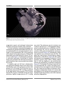
Figure 1. Computed tomography viewed on True3D Viewer (Echopixel, Inc., Mountain View, CA) showing paravalvular tunnel in the posteromedial side of the pulmonary valve.
oxygenation support and prolonged tracheostomy leading to severe post-traumatic stress syndrome.
On recent evaluation, computed tomography scan showed grossly calcified valve leaflets with a PVL tun- nel measuring 8x6 mm and approximately 9 mm in length in the posteromedial side of the pulmonary valve adjacent to the right aortic sinus. Moderate RV hypertrophy and mildly dilated RV volume (132 mL/ m2) was noted (Figure 1). He developed symptomat- ic ventricular tachycardia. Considering his complex history and associated risks with redo sternotomy, a multi-disciplinary team recommended transcatheter PVL closure.
A complete right and left heart catheterization was performed. Moderate stenosis was noted across the Mitroflow valve at the pulmonary position with gradient of 25-30 mmHg. The RV pressure was su- pra-systemic measured at 86/11mmHg as compared to the right femoral artery pressure of 74/47 mmHg. Elevated PA pressure of 56/6(28) mmHg with mean pulmonary capillary wedge pressure of 12 mmHg
was noted. The pulmonary vascular resistance was 5.2 Wu/m2 with Qp:Qs of 1:1. PA angiography demon- strated moderate pulmonary insufficiency and PVL. The pulmonary PVL site was localized by balloon occlusion of the valve and simultaneous contrast in- jection into the PA (Figure 2A). Subsequently, a 0.035 inch glidewire was positioned across the PVL. The defect was sized using an Armada balloon (Abbott, Abbott Park, IL, USA; 8x4 mm; Figure 2B). After careful hemodynamic and angiographic evaluation, a 12mm Amplatzer vascular plug II (AVP II; Abbott, Abbott Park, IL, USA) was deployed in the tunnel-like leak without any complications or significant residual leak (Figure 2C). The diastolic PA pressure improved from 6 to 20 mmHg after PVL closure. The patient recov- ered well and was discharged to home the following day. The 6-week post-procedure transthoracic echo- cardiogram showed peak velocity of 3.2 m/sec with peak gradient of 42 mmHg and mean gradient of 24 mmHg through the pulmonary valve with moderate pulmonary regurgitation.
Journal of Structural Heart Disease, February 2019
Volume 5, Issue 1:21-24
