
Page 31 - Journal of Structural Heart Disease Volume 5, Issue 2
P. 31
Case Report
44
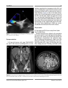
Figure 1. Transesophageal Echocardiogram of the patent fora- men ovale with color doppler.
Case presentation
A 65-year-old woman with stage T3bN1M0 RCC was seen in the structural cardiology clinic as a con-
sultation regarding the management of her PFO and severe AS before the surgical resection of the recently discovered left RCC with an extensive IVC tumor-re- lated thrombus (Figure 2). Due to the high intraop- erative risk of clot embolization causing a stroke via the PFO and the high surgical risk given her severe AS, it was decided to proceed with PFO closure and aortic valve replacement (AVR) before her radical ne- phrectomy. She was evaluated by our heart team and deemed high risk for surgical AVR, hence Transcath- eter Aortic Valve Replacement (TAVR) was recom- mended for the treatment of her aortic stenosis.
The procedure
R-IJ vein approach PFO Closure using 30 mm Cardio- form Occluder Device
General anesthesia was utilized in this procedure due to the need for transesophageal echocardiogra- phy (TEE). A 6Fr sheath was inserted into the RIJ and hemodynamic measurement was obtained show- ing the following hemodynamics: Right atrial (RA) pressure=12mmHg, systolic pulmonary artery pres- sure=33mmHg with a mean of 24mmHg, post-cap- illary wedge pressure=17mmHg, estimated cardiac
Figure 2. Computerized tomography images of the left renal mass (Panel A) and thrombus (Panel B) extending through the left renal vein into the inferior vena cava (IVC). *: Indicates the renal mass and thrombus extending into the IVC.
Journal of Structural Heart Disease, April 2019 Volume 5, Issue 2:43-47
