
Page 36 - Journal of Structural Heart Disease Volume 5, Issue 2
P. 36
49 Case Report
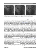
Figure 1. Initial coronary angiogram demonstrating severe stenosis of the aorta to LM “snorkeled” stent (Panel A). Subsequent coro- nary angiogram after initial stent placement (Panel B). Final coronary angiogram after second stent placement (Panel C) .
Case Presentation
We present a case of a 71-year-old woman with coronary artery disease and aortic stenosis treated 10 years previously with single-vessel coronary artery bypass (SVG- RCA) and surgical aortic valve replace- ment with a 21 mm Mitroflow for non-rheumatic aortic valve stenosis. She then presented 8 years lat- er with bioprosthetic valve degeneration and severe aortic regurgitation and underwent TAV-in-SAV with a 23 mm Medtronic Evolut R valve. Because of con- cern for left main (LM) occlusion with TAVR deploy- ment due to a low take-off of the LM (8 mm from the plane of the annulus) a 4.0 x 15 mm Xience Alpine drug-eluting stent was positioned over a 0.014 Luge guidewire prior to valve placement. PEA arrest due to LM obstruction occurred immediately with TAVR deployment, and subsequently, the stent was pulled back and deployed in a snorkel fashion from the left main to the aorta behind the side struts of the Evolut R valve with the successful return of circulation and completion of the procedure.
The patient did well for two years but developed chest pain and progressive dyspnea on exertion. A pharmacologic PET stress test provoked chest pain and demonstrated marked ischemia in the LAD and LCx distribution. She was admitted for coronary an- giography and possible intervention in the setting of the markedly abnormal stress test.
Coronary angiography was performed via right radial arterial access. The previously placed “snorkel”
stent in the LM was engaged with an EBU 3.5 guide catheter. Angiography demonstrated a 90% ostial in- stent stenosis at the location where the stent passed over the Mitroflow valve strut behind the Evolut R valve (Figure 1, Panel A). The stent and left main was wired with a Pilot 50 guidewire into the circumflex coronary artery. Intravascular ultrasound was per- formed in the LM with a Volcano Eagle Eye Platinum ultrasound catheter. The "snorkel" segment of the LM stent demonstrated a severe stenosis at the upper edge of the strut of the Mitroflow valve. The stenosis was predilated with an NC Sprinter 4.0 x 15 mm bal- loon, and a Xience Alpine 4.0 x 18 mm drug eluting stent was placed within the previous stent, extend- ing through the cell of the Evolut R into the aorta. The stent was post-dilated with an NC Quantum Apex 4.5 x 15 mm balloon. We attempted to repeat IVUS imag- ing but were unable to fully advance the IVUS cathe- ter over the Pilot 50 guide wire. The Pilot 50 wire was exchanged for a Wiggle wire through a Turnpike LP, and the IVUS catheter was able to be advanced into the stent. IVUS imaging demonstrated an ellipsoid shape to the new stent. (Figure 2, Panel A, Figure 1, Panel B) It was thought that increased radial strength was required to maintain stent patency against the external compression, and so a 4.0 x 12 mm Xience Alpine stent was placed and post-dilated with an NC Quantum Apex 5.0 x 12 mm balloon, inflated to 20 atmospheres. IVUS was repeated at the LM “snor- kel” stent segment and demonstrated an improved, less ellipsoid geometry with an MLD of 5.0 x 3.5 mm
El-Haddad H. et al.
Repeat PCI of a Post TAVR Left Main Snorkel Stent
