
Page 101 - Journal of Structural Heart Disease Volume 5, Issue 4
P. 101
163
Meeting Abstracts
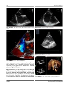
133. Figure 1. TEE demonstrates PFO, redundant Eustachian valve.
133. Figure 2. Rt. to Lt. shunt through PFO.
via an extravenous puncture to prevent its interference with the device . The PFO had been closed with no resid- ual shunt and no complications . TCD repeated revealed absence of right to left shunt.
Discussion: Most cases of right to left shunting through a PFO are seen where right atrial pressure exceeds that of the left, . But right to left shunt can be observed even if the pressure is equal, it had been reported that dilated ascending aorta may induce flobby aneurysmal septum So, we suggest that aortic root dilatation may have a role in
133. Figure 3. Inter-atrial septal aneurysm.
133. Figure 4. Dilated aortic root.
133. Figure 5. Three dimensional TEE demonstrates PFO.
Hijazi, Z
22nd Annual PICS/AICS Meeting
