
Page 18 - Journal of Structural Heart Disease Volume 5, Issue 4
P. 18
Meeting Abstracts
80
A CASE OF DEXTROCARDIA WITH KARTARGENER SYNDROME
Neeraj Awasthy
Max Hospital, Saket, Delhi, India
An 8 year old child with 10 mm non restricted vsd with dextrocardia and Kartageners syndrome was found not suitable for closure in view of marked lung issues, bron- chospasm and repeated chest infection. Patient under- went percutaneous vsd device closure through antegrade approach using 10/12 duct occluder device with signif- icant resolution of symptoms. We postulate that in view of decease lung segment, rest of the lung segment get flooded with the flow through the shunt lesions and com- promise lung compliance. The closure of defects in such sit- uations greatly facilitates the symptomatic improvement.
13. ISOLATED CONGENITAL LEFT CORONARY ARTERY
TO CORONARY SINUS FISTULA IN A NEONATE
William Fogarty1, Robert English1,2
1University of Florida College of Medicine Jacksonville, Jacksonville, USA. 2Wolfson Children's Hospital, Jacksonville, USA
Case Report: A 20-year-old presented for her first prena- tal care visit at 32 weeks gestation. Her pregnancy was uneventful. Past medical history was remarkable for schizo- phrenia and anemia. She denied use of medications, alco- hol, tobacco, and illicit substances. Family history was unremarkable. The initial fetal ultrasound demonstrated an enlarged heart and aortic narrowing.
Fetal echocardiograms at 36 and 38 weeks gestation sug- gested: total anomalous pulmonary venous return with obstruction of the pulmonary venous confluence in the left atrium, right sided chamber dilation, right ventricular pressure and volume overload, and aortic flow reversal.
Because of the likelihood of severe pulmonary hyperten- sion and obstructed pulmonary venous return after birth, a C-section was performed at 39 weeks gestation.
The child’s APGAR scores were 8 at 1 and 5 minutes. She was responsive and alert with no respiratory distress but was intubated due to the suspected cardiac pathology. Cardiovascular exam noted a soft systolic murmur to the left sternal border. An echocardiogram revealed normal pulmonary venous return and a severely dilated left main coronary artery. There appeared to be a fistula connecting the left coronary system to the coronary sinus.
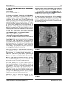
Subsequent coronary angiography revealed the fistu- lous connection to the coronary sinus off of the distal left
circumflex coronary artery. Embolization of the fistula was performed using 2 coils (14cm x 4 mm distally and 14 cm x 6 mm proximally). Repeat coronary angiography con- firmed complete occlusion of the fistula and good filling of the left coronary artery. See Figures 1 and 2.
The infant remained stable and was started on aspirin. Serial echocardiograms demonstrated closure of the PDA, persistent large atrial septal defect, aneurysmal atrial sep- tum, and normal biventricular function. The infant was dis- charged on day of life 19.
13. Figure 1
13. Figure 2
Journal of Structural Heart Disease, August 2019
Volume 5, Issue 4:75-205
