
Page 74 - Journal of Structural Heart Disease Volume 5, Issue 4
P. 74
Meeting Abstracts 136
97. TRANSCATHETER MECHANICAL MANIPULATION inflammatory material. In the setting of his life-threaten- OF OBSTRUCTED ST. JUDE PROSTHETIC MITRAL ing pulmonary hemorrhage, all anticoagulation was held. VALVE IN AN INFANT After interdisciplinary discussions, the patient was deemed
Yousef Arar, Jeff Hong, Thomas Zellers, Surendranath R. Veeram Reddy
University of Texas Southwestern, DALLAS, USA
Prosthetic valve thrombosis (PVT) is a serious complication of prosthetic heart valves that leads to leaflet immobility and ultimately valve dysfunction. Management of PVT is with either surgical intervention or systemic thrombolysis, but in patients with contraindications to both treatment modalities, options can be limited. We describe a novel approach to treat prosthetic valve obstruction and leaflet immobility in an infant who was a very poor candidate for both surgery and systemic thrombolysis. To our knowl- edge, this is the youngest patient to undergo prosthetic mitral manipulation.
Case Report: An ex-35 weeks gestational age male with congenital arcade mitral valve underwent a surgical mitral valve replacement with a 17 mm St. Jude aortic mechan- ical prosthesis at 9 months of age. At 13 months of age he presented with acute respiratory distress and hemop- tysis, with bronchoscopy showing active bleeding in his right lower lung. Bedside fluoroscopy confirmed that the medial mitral valve leaflet was completely immobile, most likely secondary to obstruction by adherent fibrinous and
a very poor candidate for systemic thrombolysis and car- diac surgical therapies.
From a right femoral vein approach and 6-French (Fr) per- cutaneous access, a transseptal puncture was performed to gain access to the left atrium. Attention was focused on using catheters to manipulate the immobile leaflet of the mechanical mitral valve. Using TEE and fluoroscopic guid- ance, the medial leaflet was struck repeatedly with multi- ple catheters (4 Fr angled glide, 4 Fr 3.0 curve Judkins Right (JR), and 5 Fr 4.0 curve JR catheters) with no significant improvement in leaflet excursion. The 4.0 curve of the 5 Fr JR catheter was then reinforced with a 0.035” tip deflect- ing wire (TDW) that enabled the catheter to eventually be advanced across the immobile leaflet from the left atrium into the left ventricle. The catheter with the TDW in situ was then withdrawn from the left ventricle to position it across the medial hinge points of the valve leaflet and gen- tle force was applied to push open the valve completely. Post-intervention, there was no residual gradient across the mitral valve (LAP = 17, LVEDP = 18). A catheter was then placed in the left atrium for local tPA infusion to prevent further thrombosis.
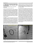
97. Figure 1. Catheter tip across the St. Jude valve into the left ventricle status post catheter manipulation.
Journal of Structural Heart Disease, August 2019 Volume 5, Issue 4:75-205
