
Page 13 - Journal of Structural Heart Disease - Volume 1 Issue 1
P. 13
7
Original Research Article
Table 1. Adopted from Holmes et al. [6]
Table 2. TAVR Follow-Up AQ6
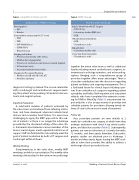
Components of TAVR Screening
Demographics • Age
• Gender
Comorbidities (many used for STS Score) • CAD
• PVD
• CHF (NYHA Class) • COPD (FEV1)
• Renal Function
Imaging to Confirm
• Presence and severity of AS (echo)
• CAD burden (angiography)
• Presence of cerebral vascular disease (carotid doppler) • LV Function (echo)
• Associated valvular lesion (echo)
Imaging for Procedural Planning
• Annular size (2D and 3D echo, CT)
• Aortoiliac anatomy
Post-Procedure Follow Up
Initial 2-Week Visit with CT Surgery • Chest X-ray
• Laboratory studies (BMP, etc.) • ECG
1-Month Visit in Valve Clinic • ECG
• Transthoracic echo
6-Month Visit
• Laboratory studies only
1-Year Visit then Yearly Visits • Transthoracic echo
diagnostic testing is planned. The session culminates with a cardiologist and cardiothoracic surgeon meet- ing the patient and presenting all medical, interven- tional, and surgical options.
Inpatient Evaluation
together the entire valve team as well as additional faculty including several cardiothoracic surgeons, in- terventional cardiology operators, and echocardiog- raphers. Bringing such a comprehensive group of providers together offers many advantages. There is a broader contribution into the discussion regarding patient candidacy and ongoing management. This is a facilitated forum for clinical input including opin- ions from cardiothoracic surgeons regarding patient operability and frailty. Both inpatient and outpatient referrals who have completed the extensive screen- ing for TAVR or MitraClip (Table 1) are presented. Im- portantly, this is also an opportunity to prioritize and schedule patients for procedure allowing timely de- livery of care to those most in need of treatment.
Follow Up
Post procedure patients are seen initially at 2 weeks by cardiothoracic surgery at which time they undergo evaluation with chest X-ray, electrocardio- gram, and laboratory studies (Table 2). Subsequently, patients are seen in valve clinic at 1 month, 6 months, 12 months, and then yearly thereafter. Echocardio- graphic studies are obtained prior to discharge, 1 month and then yearly. Again, having the MDT avail- able at valve clinic provides the ability to address a wide range of post-procedural issues.
A substantial number of patients evaluated by the valve team are transferred from referring institu- tions and have developed advanced valvular heart disease and secondary heart failure. It is even more challenging to apply the MDT approach to this sub- set of patients as there is no longer the advantage provided by the structured setting of the valve clin- ic. Communication between various members of the team is crucial. Again, a well-organized central core of support staff can facilitate this task and help meet the goal of patient evaluation by both a CT surgeon and cardiologist within 24 hours.
Weekly Meeting
Complementary to the valve clinic, weekly MDT meetings are held at our institution. This weekly valve meeting is the nucleus for the valve service and brings
Lasala, J.L. et al.
Building a Structural Heart Disease Team
