
Page 32 - Journal of Structural Heart Disease - Volume 1 Issue 1
P. 32
Original Research Article
26
more manageable. The basic technique involves obtaining femoral arterial access, performing suture preclosure, and placing an arterial sheath ranging in diameter from 10 up to 14 French, depending on the balloon type and size. Meticulous technique using fluoro- or ultrasound-guided femoral access to ensure sheath insertion in a non-calcified segment of the common femoral artery is critical (Figure 2). After access is obtained, the valve is crossed using standard techniques (Figure 3) and stiff exchange wire is placed as a rail for delivery of the balloon. The balloon is advanced retrograde over the guidewire into the left ventricle and positioned across the native aortic valve annulus. Rapid pacing at a heart rate, typically between 170 and 200 beats per minute is initiated and when the blood pressure falls, the balloon is inflated (Figure 4). After full inflation is achieved, the balloon is withdrawn and rapid pacing discontinued. If the balloon was ejected from the ventricle during the inflation, and blood pressure recovery has been adequate, a second inflation may be performed. At that point, the result is assessed and if appropriate to the situation, a larger balloon can be used when the result does not meet the planned expectations. The balloon and wire are withdrawn, and the existing suture is used to close the arterial sheath insertion site, usually with a wire left in place so that if the pre- closure fail, an additional Perclose device can be used or a sheath inserted so that manual compression may be undertaken when the anticoagulation normalizes or is reversed. Careful attention to the hemodynamics before and after each inflation is crucial. While it is not necessary to re-measure the transaortic gradient after each inflation it is critical to carefully assess the changes in aortic diastolic pressure as an indicator of increase aortic insufficiency. If a second inflation is necessary, the possibility of more than mild aortic insufficiency should be excluded before proceeding for the second attempt.
Antegrade BAV
A much less utilized approach is to deliver a bal- loon into the native aortic annulus using transve- nous-trans-septal access [11]. From the trans-septal puncture into the left atrium, a single-lumen bal- loon catheter can be floated into the left ventricle
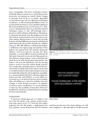
Figure 8.
The left femoral artery access can be used to intro- duce a 10mm gooseneck snare to catch the distal end of the 0.032 inch wire.
Figure 8 Video.
and then into the aorta. This allows delivery of a stiff wire antegrade so that the balloon for BAV may be
Structural Heart Disease, May 2015
Volume 1, Issue 1: 20-32
