
Page 33 - Journal of Structural Heart Disease - Volume 1 Issue 1
P. 33
27
Original Research Article
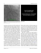
Figure 9. The snared support wire may be left parked in the descending aorta, as this will provide adequate support for ante- grade balloon passage.
introduced on the venous side of the circulation and tracked through the left atrium and left ventricle to straddle the aortic valve. Antegrade BAV is technical- ly more demanding than retrograde BAV. Many of the procedure steps are unfamiliar to many BAV and TAVR operators. One of the advantages of antegrade BAV is utilization of a vein rather than an artery for access. This is occasionally useful in patients with se- vere peripheral arterial disease. Another advantage is stability of the balloon during inflation in the aor- tic valve even without rapid pacing. Since there is an arterial-venous loop, the balloon can be controlled from both antegrade and retrograde directions and is highly stable. In addition, the use of venous access allows for the utilization of much larger diameter bal- loons. In our series, this resulted in larger acute valve areas BAV compared to a retrograde technique.
The next several paragraphs will detail the specific procedure steps of antegrade BAV. The initial set up for antegrade BAV includes 7 French left femoral arte- rial and 7 or 8 French left femoral venous access, and for the trans-septal puncture, 14 French right femoral venous access. The arterial access sheath size is re- quired to place a 10mm gooseneck snare via the left
Figure 9 Video.
femoral arterial sheath and still have enough room for arterial pressure measurement. The left femoral venous access is for initially pulmonary artery cath- eterization and cardiac output measurement, and subsequently for medication administration if need- ed. The 14 French right femoral venous access facili- tates trans-septal puncture and allows for placement of a Inoue balloon catheter.
After access is obtained, trans-septal puncture is performed. A posterior mid fossa level puncture al- lows easiest access to the mitral orifice. A 7 French single-lumen balloon catheter is inflated in the left atrium and with counterclockwise rotation of a stan- dard Mullins sheath the balloon tip catheter is float- ed across the mitral valve (Figure 5). At this point, the transaortic valve pressure gradient can be mea- sured. A standard 0.035” J-tip wire can be introduced into the single-lumen balloon catheter with a large curve added, so that the balloon catheter can be di- rected around the left ventricular apex (Figure 6). At this point, it is sometimes necessary to switch to a floppy tip straight wire such as a Wholey wire to cross the aortic valve antegrade and with the single-lumen catheter balloon deflated, pass the balloon catheter
Feldman, T. et al.
Balloon Aortic Valvuloplasty
