
Page 35 - Journal of Structural Heart Disease - Volume 1 Issue 1
P. 35
29
Original Research Article
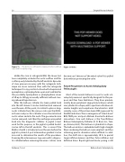
Figure 11. The balloon is inflated in stepwise fashion without any need for rapid pacing.
Unlike the case in retrograde BAV, the Inoue bal- loon completely occludes the aortic outflow and this is often poorly tolerated by the left ventricle. Episodes of slow pressure recovery with the antegrade tech- nique are more common than with the retrograde technique. It is my practice to have both atropine and epinephrine or phenylephrine open and available for this possibility. Epinephrine or phenylephrine doses of 50 up to 250 μg are usually sufficient without caus- ing rebound hypertension.
After the balloon catheter has been pulled back into the left atrium it can be stretched and removed over the wire. At this point, it is critical to place a diag- nostic catheter from the venous side over the 0.032- inch wire and pass this catheter across the mitral and aortic valves and into the aorta. The gooseneck snare can be removed, and then the exchange wire pulled back into the diagnostic catheter. A pigtail is best used for this purpose, as the pigtail is pulled back it can be left in the left ventricle. This is easier if the Mullins sheath is introduced over the wire before the pigtail is placed. A post intervention gradient can be assessed to determine the results of the procedure. Covering the arteriovenous loop wire with a diagnos- tic catheter before removing the wire is critical, since
Figure 11 Video.
the wire can "cheese cut" the mitral valve if it is pulled back without protecting the valve.
Future Perspectives in Aortic Valvuloplasty Technologies
Most of the current balloons in use for aortic val- vuloplasty were not specifically designed for this pur- pose and thus have limitations. They have predom- inantly been peripheral angioplasty balloons which are cylindrical in shape, with a spectrum of balloon di- ameter lengths and compliance characteristics. Until recently, none of these have been FDA approved for the BAV indication. The re-emergence of“stand-alone” BAV, TAVR pre- and post-dilations have led to balloon innovations. One such balloon is the True Dilation Balloon Valvuloplasty Catheter (C. R. Bard, Inc., Tem- pe, Arizona, USA). This balloon is cylindrical in shape. The balloon matrix is embedded with high-strength fibers rendering the balloon noncompliant and thus achieving precise diameters when inflated. In addi- tion, the shaft size is approximately 10 Fr to permit larger inflation lumens and thus faster inflation and deflation times. Rapid right ventricular pacing is still
Feldman, T. et al.
Balloon Aortic Valvuloplasty
