
Page 21 - Journal of Structural Heart Disease Volume 1, Issue 3
P. 21
Original Scientific Article
Journal of Structural Heart Disease, August 2015, Volume 1, Issue 3: 127-136
DOI: http://dx.doi.org/10.12945/j.jshd.2015.018-14
Received: December 24, 2014 Accepted: January 16, 2015 Published online: August 2015
An Overview of the Mitraclip Procedure
Indications, Procedural Characteristics, and Clinical Outcomes
Rahul P. Sharma, MD*, Moody Makar, MD, Saibal Kar, MD Cedars Sinai Heart Institute, Los Angeles, California, USA
Abstract
The MitraClip procedure is a safe and effective ap- proach to reduction of mitral regurgitation (MR) with proven durability and clinical improvement. Procedur- al success is dependent on patient selection, under- standing of mitral valve anatomy, particularly from an echocardiographic perspective, and attention to crit- ical elements of the implantation such as trans septal puncture.
In the United States, the FDA has approved the Mi- traClip device for treatment of high risk patients with primary MR. The question of long term, sustained re- duction of MR and persistent clinical improvement re- mains to be addressed with longer duration of follow up. Based on the impeccable safety profile of the pro- cedure and demonstrated medium term clinical dura- bility, future studies should be aimed at the evaluation of MitraClip for treatment of patients with severe MR deemed moderate, or indeed low risk, for surgery. Copyright © 2015 Science International Corp.
Key Words
MitraClip • Mitral • Regurgitation • Valve
Anatomical Considerations
An understanding and appreciation of the com- plex anatomy of the mitral valve (MV) apparatus is imperative to achieving procedural success with the MitraClip (Abbott Vascular, Santa Clara, California, USA) device.
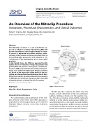
Figure 1: Mitral anatomy
The MV apparatus comprises the mitral valve, the annulus, annular attachment at the atrio-ventricular junction, tendinous chords, and the papillary mus- cles. The valve is made up of two leaflets, commonly referred to as the anterior and posterior leaflets (oc- casionally referred to as the mural and aortic leaflets, respectively). The posterior leaflet is narrow compared to the anterior leaflet and extends two-thirds around the left atrio-ventricular junction within the inlet por-
* Corresponding Author:
Saibal Kar, MD
Cardiovascular Intervention Center Research
Cedars-Sinai Medical Center
Heart Institute, Los Angeles 90048, USA
Tel.: +1 310 423 3977, Fax: +1 310 423 0106, Email: Saibal.Kar2@cshs.org
Fax +1 203 785 3346
E-Mail: jshd@scienceinternational.org http://structuralheartdisease.org/
© 2015 Journal of Structural Heart Disease Published by Science International Corp. ISSN 2326-4004
Accessible online at:
http://structuralheartdisease.org/
