
Page 22 - Journal of Structural Heart Disease Volume 1, Issue 3
P. 22
Original Scientific Article
128
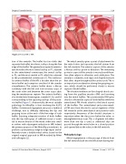
Figure 2: Primary MR vs FMR
tion of the ventricle. The leaflet has two clefts that separate the leaflet into three scallops along the free edge of the leaflet. The generally accepted nomencla- ture describes the most lateral scallop as P1, adjacent to the anterolateral commissure, the central scallop as P2, and the most medial as P3, which lies adjacent to the posteromedial commissure [1]. The semicircu- lar anterior leaflet of the MV is broader than the an- terior leaflet and comprises one-third of the annular circumference. The anterior leaflet shares a fibrous continuity with the left and non-coronary cusps of the aortic valve and between the aortic cusps abut- ting the membranous septum. The anterior leaflet is also divided into three regions, namely A1, A2, and A3 corresponding to the opposing scallops of the poste- rior leaflet (Figure 1). Anatomically, the most suitable pathology for MitraClip is that involving the A2/P2 leaflets. Commisural regurgitant jets pose a technical challenge, due to difficulty delivering the clip and grasping tissue at the ends of the free edge of each leaflet. Ensuring adequate insertion of both leaflets into the clip with grasp of sufficient tissue is essen- tial to ensure division of the mitral orifice into small- er orifices with subsequent reduction in MR. Indeed, the primary purpose of the MitraClip procedure is to perform a percutaneous edge-to-edge repair and ef- fectively create a double mitral orifice, based on the original surgical approach to MR described by Alfieri and colleagues [2].
The mitral annulus gives a point of attachment for the mitral valve and separates the left atrium from the left ventricle. The anterior aspect of the annulus is fibrous and less prone to dilatation. The remaining posterior aspect of the annulus is muscular and there- fore often subject to dilatation and calcification. The annulus is a dynamic, non-rigid, oval shaped structure that alters shape throughout the cardiac cycle. This is an important consideration during the grasping pro- cess, which should be performed slowly to ensure capture of both leaflets.
The chordae tendinae are fan-shaped chords aris- ing from the papillary muscles (PM) and inserting into the mitral leaflets. The posteromedial PM gives chords to the medial aspect of both leaflets while the anterolateral PM chords attach to the lateral aspect of the leaflets. The anterolateral and posteromedi- al PM arise from the mid to apical segments of the left ventricle at the anterolateral and posterior walls respectively. Awareness of the chordal structures is important when the clip passes below the valve, as entanglement may occur. This is of greater risk when more than one clip is used, as additional clips are passed through the mitral valve in a closed position and opened below the valve, in the left ventricle.
Pathophysiology
Mitral regurgitation is the passage of blood from the left ventricle back into the left atrium during ven-
Journal of Structural Heart Disease, August 2015
Volume 1, Issue 3: 127-136
