
Page 14 - Journal of Structural Heart Disease Volume 1, Issue 4
P. 14
159
Meeting Abstracts
deployed at the base of the LAA, achieving complete occlusion. The patient tolerated the procedure well, and following an uneventful postoperative course he was discharged to home 3 days later. Fol- lowup gated cardiac CT angiography showed complete closure of the LAA by Atriclip-Pro.
Discussion and Learning Points: Device-assisted epicardial closure of the LAA is an evolving option for the prevention of systemic embo- lism in patients with AF. While this is a safe and e ective therapy for patients who are intolerant of anticoagulation, there is limited experi- ence with these devices, and operators need to be aware of the poten- tial for both early and delayed complications. To our knowledge, this is the rst reported case of the use of a thoracoscopically deployed Atriclip-Pro device to acutely salvage an incomplete LAA ligature by the LARIAT device.
During deployment, LAA anatomy was such that, even with incom- plete closure of the LAA by both the LARIAT and the LARIAT Plus liga- tures, the appendage appeared occluded by direct compression from the LARIAT suture delivery device. After the LARIAT delivery catheter was removed o the LAA base and angiography was again performed, a remaining trabeculated infundibulum was noted. In contrast to the smooth-walled residual “stump” which is sometimes seen following epicardial LAA exclusion, this remaining trabeculated portion of the LAA may serve as a potential cardioembolic source. Furthermore, early thrombosis at the site of LAA ligation has been well described following the LARIAT procedure, and is postulated to be the result of endothelial injury and in ammation producing a nidus for thrombus formation. Due to the residual risk of LAA thrombosis, the patient was referred to cardiac surgery service for completion of LAA exclusion.
Although totally thoracoscopic deployment of the Atriclip-Pro device is well described, its use in this scenario is novel. This case demon- strates the feasibility of completion of LAA closure following incom- plete LAA ligation by sub-xiphoid approach. Additionally, this case highlights the possibility of incomplete LAA closure despite a favor- able appearance on angiography during deployment of the LARIAT suture delivery device.
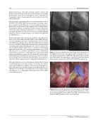
Figure 1. Intra-procedural uoroscopy during initial deployment of the LARIAT over the neck of the LAA (a), with incomplete closure on post ligation angiogram (b). LARIAT Plus deployment, again over the neck of the LAA (c). Final angiographic appearance of the LAA, with the trabeculated secondary lobe una ected by LARIAT Plus ligature (d).
Figure 2. Thoracoscopic appearance of the Atriclip device (AC, white), over the previously ligated left atrial appendage (LAA, blue) as well as the previously una ected secondary lobe (LAA*, yellow). Previously deployed LARIAT ligatures are also seen (Lig, black).
CSI Africa 2015 Meeting Abstracts
