
Page 22 - Journal of Structural Heart Disease Volume 2, Issue 1
P. 22
Original Research Report
16
Video 1. Subcostal bicaval view, documenting adequacy of superior vena caval (SVC) and inferior vena caval (IVC) mar- gins. View supplementary video at http://dx.doi.org/10.12945/j. jshd.2016.007.14.vid.01.
Video 2. Skewed Apical 4 chamber view with color compare showing the atrio-ventricular (or mitral) margin and the posterior margin. View supplementary video at http://dx.doi.org/10.12945/j. jshd.2016.007.14.vid.02.
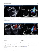
Slide # 14:
Left frame: Subcostal bicaval view, documenting adequacy of superior vena caval (SVC) and inferior vena caval (IVC) margins (Video 1).
Right frame: Skewed Apical 4 chamber view with color compare showing the atrio- ventricular (or mitral) margin and the posterior margin (Video 2).
Video 3. TEE at 0 degrees (4-chamber view), showing the AV valve (or mitral) and the posterior margins. View supplementary video at http://dx.doi.org/10.12945/j.jshd.2016.007.14.vid.03.
Video 4. TEE at 40 degrees (aortic short axis view), depicting the retroaortic and the posterior margins. View supplementary video at http://dx.doi.org/10.12945/j.jshd.2016.007.14.vid.04.
Slide # 16:
TEE imaging in di erent views to con rm adequa- cy of surrounding margins for device closure of the ASD.
Top left: TEE at 0 degrees (4-chamber view), show- ing the AV valve (or mitral) and the postero-inferior margins (Video 3).
Journal of Structural Heart Disease, February 2016
Volume 2, Issue 1:15-32
