
Page 42 - Journal of Structural Heart Disease Volume 2, Issue 1
P. 42
Original Research Article
36
Introduction
ciated with complex heart lesions, and or contraindication to antiplatelet therapy.
Hospital IRB approval for the study was obtained. Data col- lected included patient’s demographics, echocardiographic and cardiac catheterizations data (Tables 1 and 2). Electrocar- diographic (EKG) data was collected and analyzed to evaluate the rhythm disorders in this cohort (Tables 3 and 4).
Previously described technique for VSD closure was used [14]. Echocardiogram and EKG on the rst post-catheteriza- tion day and at 1, 3, 6, and 12 months, and then yearly were reviewed for any conduction abnormality, residual VSD shunt, aortic regurgitation (AR), or tricuspid regurgitation (TR).
The data were initially entered into an Excel spread sheet and subsequently imported into the JMP Statistics Package v8.0.1 (SAS Corp., USA). All statistical analyses were carried out within JMP. In addition to basic descriptive statistics (mean, medians, ranges, standard deviations, and counts of missing data), both Pearson’s and Spearman’s correlation coe cients (and their associated p values) were calculated to explore the linear correlations between speci c pairs of variables, and the correlations quoted here are Spearman’s rho. Potential outli- ers were identi ed by eye (based on JMP scatter plots) and aided by the superimposition of a 95% bivariate normal den- sity ellipse generated by JMP. The statistical signi cance of the di erences between the means of continuous variables was explored using the t test (for equal or unequal variances as ap- plicable) and Wilcoxon’s test, and the p values quoted here are for Wilcoxon’s. The alpha-level for statistical signi cance was set to be 0.05 for all tests.
Results
VSD device closure was attempted on 49 patients with successful closure achieved in 45 patients (success
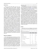
Table 1: Patient demographics, VSD size, device size, Qp:Qs, RVSP, uoro time, and procedure time
Ventricular septal defect (VSD) is the most com- mon congenital heart disease (CHD), constituting 30–40% of all congenital heart diseases [1]. Symptom- atic patients need medical management and probably surgical closure if medical therapy fails. Although ad- vances in surgical, anesthetic, and postoperative care have made surgical closure of VSD safer, morbidities like cerebrovascular accidents, seizures, chorea/athe- tosis, lung collapse, phrenic nerve injury, and junc- tional ectopic tachycardia are still concerns following open heart surgery [2, 3]. One of the serious compli- cations of surgery is complete atrioventricular block (CAVB), which has been reported from 0.7% to 3.1% for membranous and outlet VSDs [4, 5]. Transcatheter approach to close such VSDs has been an attractive option to avoid these morbidities. Ten years after the initial transcatheter closure, in 1998, the Amplatzer muscular occluder had revolutionized the percutane- ous VSD closure with favorable outcomes [6, 7]. Ap- proximately two-thirds of VSDs are in the perimembra- nous (pmVSD) location, and there have been growing concerns about complete heart block at late follow up of percutaneous membranous VSD closure. A special Amplatzer membranous VSD device with an eccentric left ventricular disc was designed for the closure of pmVSDs with good initial results [8–10]. Occasionally, pmVSD with aneurysmal tissue can be closed with an Amplatzer duct occluder (ADO) [11, 12]. The aim of this study is to evaluate rhythm disturbances caused by transcatheter VSD devices at immediate and long-term follow up.
Materials and Methods
This is a retrospective observational study to assess imme- diate to long-term rhythm follow up of percutaneous closure of muscular VSD (mVSD) and pmVSD with di erent types of Amplatzer occluders. All patients who were taken to the cath- eterization laboratory for attempted VSD closure using an Amplatzer device during the period of January 2003 and Sep- tember 2012 at Hamad General Hospital, Doha, Qatar, were included. Inclusion criteria for percutaneous VSD closure was a muscular or pmVSD with clinical and or echocardiographic evidence of signi cant left to right shunt or a signi cant re- sidual VSD after surgical repair. Exclusion criteria for percu- taneous VSD closure was a body weight less than 7 kg, right to left shunt across the VSD, a pmVSD with less than 2-mm subaortic rim on long axis echocardiographic view, VSDs asso-
Age (years)
Weight (kg)
VSD size (mm) by TTE
VSD size (mm) by TEE
VSD size (mm) by LV angiography Device size (mm)
Qp:Qs
RVSP (mm Hg)
Fluoro time (min)
Procedure time (min)
11.2 8.5 35.5 24 7.2 7
7.5 6
6.2 6
8.7 8
1.5 1.4 31.5 28.5 47.11 41.5 145 132.5
2–36.7 10–106 3–14 4–15 3–14 4–18 1– 3 20–50 17–138 46–310
Mean
Median
Range
TTE = transthoracic echo; TEE = transesophageal echo; Qp:Qs = pulmonary to systemic blood ow ratio; RVSP = right ventricular systolic pressure.
Journal of Structural Heart Disease, February 2016
Volume 2, Issue 1: 35-41
