
Page 43 - Journal of Structural Heart Disease Volume 2, Issue 1
P. 43
37
Original Research Article
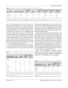
Table 2: Comparison of muscular and membranous VSD groups and their mean values
Type of VSD
Age (years)
Wt (kg)
VSD TEE (mm)
Qp:Qs
Device Size (mm)
Proc. Time (min)
Fluoro Time (min)
Follow Up (months)
Muscular Membranous P value
13.2 46.5 6.67 1.65 12.43 36.35 6.66 1.4 0.31 0.11 0.49 0.11
8.25 8.14 0.71
159.16 61 137.81 43.93 0.67 0.21
54.78 64.03 0.51
Wt = weight; TEE = transesophageal echo; Qp:Qs = pulmonary to systemic blood ow ratio; Proc time = procedure time.
rate of 91.8%); the female to male ratio was 1.14. Among these 45 VSD closure cases, 35 were perimem- branous (78%) (34 native and 1 surgical residual) and 10 muscular (9 native and 1 surgical residual) defects. The pmVSDs were closed using 23 pmVSD devices, 6 muscular VSD devices, and 6 Amplatzer duct occluders (ADO), whereas the muscular VSDs were closed with nine muscular VSD devices and one ADO device. The ADO devices were used only in tunnel shape aneurysmal VSDs. Ten (22%) patients had asso- ciated cardiac anomalies including patent ductus ar- teriosus in one, pulmonary valve stenosis in four, mild mitral stenosis in one, additional small muscular VSDs in two, and bicuspid aortic valve in two.
In two out of four unsuccessful cases, the VSD de- livery sheath could not be advanced through VSD due to aneurysmal tissue; in one case, there was device
Table 3: New onset rhythm disorders in the membranous VSD group
related aortic regurgitation and the device was re- trieved with no residual regurgitation, and in another case, there was transient complete heart block that reverted to normal sinus rhythm after device remov- al. All four patients who failed device closure were re- ferred for surgical closure.
At the time of VSD closure, the median age of the study group was 8.5 years (range 2–36.7 years) and the median weight of 24 kg (range 10–106 kg). The median VSD size by transthoracic echocardiogram (TTE), TEE, and by LV angiogram was 7, 6, and 6 mm, respectively, and the device size was with a median of 8 mm (range 4–18). The median ratio of systemic to pulmonary blood ow (Qp:Qs) was 1.4 (range 1–3) and the median right ventricular systolic pressure was 28.5 mm Hg (range 20–50 mm Hg). The median uoro and the median procedure times were 41.5 minutes and 132.5 minutes, respectively (Table 1).
There was no signi cant correlation between weight and VSD size with uoroscopy time [when outliers were removed the correlation coe cient was 0.282 (p=0.11) and 0.066 (p=0.73), respectively].
Table 4: New onset rhythm disorders in the muscular VSD group
ICRBBB - 1 10.5 37 6 6 CLBBB - 1 3.5 23 6 6 PVCs - 1 8 21 4 4
TEE = transesophageal echo; CLBBB = complete left bundle branch block; ICRBBB = incomplete right bundle branch block; PVCs = premature ventric- ular contractions.
Rhythm Disorder & Patient Number
Age (years)
Weight (kg)
VSD Size (mm) by TEE
Device Size (mm)
CRBBB - 4 1
2
3
4
CLBBB - 1 CAVB - 1 EAT - 1
V. Tach - 1
17.5 57 12 12 9.4 18 14 16 5 18 8 10 5.7 14 4 4
5 16 6 6
Rhythm Disorder & Patient Number
Age (years)
Weight (kg)
VSD Size (mm) by TEE
Device Size (mm)
5 17 12 27.2 54 4 14.3 77 11
12 4 10
TEE = transesophageal echo; CRBBB = complete right bundle branch block; CAVB = complete atrio-ventricular block; EAT = ectopic atrial tachycardia; V. Tach = ventricular tachycardia.
Dilawar, M. et al.
Rhythm disturbances after device closure of VSD
