
Page 50 - Journal of Structural Heart Disease Volume 2, Issue 6
P. 50
277
Meeting Abstracts
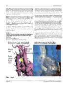
Methods: All patients with models printed from RA were selected. RA acquisitions from a Toshiba In nix-I system were postprocessed and printed with a Stratasys Eden 260. Two independent observers mea- sured 5-10 points of interest on both the RA and the 3D model. Bland Altman plot was used to compare the measurements on rotational angiography to the printed model.
Results: Models were printed from RA in 5 patients (age 2 mo - 1 yr). Diagnoses included a) coronary artery aneurysm, b) Glenn shunt, c) coarctation of the aorta, d) Tetralogy of Fallot with MAPCAs, and e) pulmonary artery stenosis. There was no signi cant measurement di erence between RA and the printed model (r= 0.990, p<0.01, Bland Altman p=0.987). There was also no signi cant interobserver variability. The MAPCAs model was referenced by the surgeon intra- operatively and was accurate.
Conclusions: Rotational angiography can generate highly accurate 3D models in congenital heart disease, including in small vascular structures. These models can be extremely useful in patient evalua- tion and management.
#0088
TRANSCATHETER DEVICE CLOSURE OF CONGENITAL LEFT SUBCLAVIAN ARTERY TO INNOMINATE VEIN FISTULA
Jessica Nikitczuk, Barry Love
Mount Sinai Medical Center, New York, NY, USA
History: A 5 week old male presented in congestive heart failure at 2 weeks of age with a loud continuous murmur. Echocardiogram and CT angiogram showed a stula between the left subclavian artery and a severely dilated innominate vein. All cardiac chambers were dilated and there was systemic-level right ventricular pressure.
Procedure: Cardiac catheterization con rmed systemic pulmonary artery pressure with a Qp/Qs (e ective) of 4:1. Angiography in the left subclavian artery demonstrated the stula measuring 2.9mm at the narrowest point. The stula was crossed from the venous side using a 4Fr Berenstein catheter and 0.018 HiTorque wire. A 6/4 Amplatzer Duct Occluder was then delivered from the venous approach placing the “hat” in the left subclavian artery and the “stem” in the innomi- nate vein. There was no residual ow after device placement and no obstruction of the innominate vein or subclavian artery. Immediately following closure, the pulmonary artery pressure fell to 1/3 sys- temic. At 6 month follow-up, the infant is thriving on no medication. Echocardiogram shows no residual ow across the stula, normal biventricular size and function and equal blood pressures in both arms.
Discussion: Congenital arteriovenous stula (AVF) is a rare cause of congestive heart failure in infancy and mainly con ned to the head and liver - only a handful of case reports describe intrathoracic AV stulas. Once identi ed, transcatheter occlusion was an e cient and e ective therapy leading to immediate resolution of heart failure and pulmonary hypertension.
Figure 1 (#0089).
Hijazi, Z
20th Annual PICS/AICS Meeting Abstracts
