
Page 23 - Journal of Structural Heart Disease Volume 3, Issue 1
P. 23
Original Scienti c Article
16
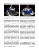
Figure 2. Transesophageal echocardiogram demonstrating a de- ployed occluding device in one of the atrial septal defects. A leak is present outside the perimeter of the device.
one of the central defects failed to occlude all open- ings (Figure 2). A second attempt was performed in the adjacent central defect using a 21-mm occluder. Residual leak was demonstrated only within the pe- rimeter of the device (Figure 3). At this stage, the de- vice was released.
The transcatheter closure of a multiple or fenestrat- ed ASD can be accomplished by several methods [2]. The defects can be closed by the use of several devic- es, with each implanted to close one or more defect. When the distance between the ASDs is greater than 7 mm, placement of two devices is recommended [3]. Closure of the larger defect should be performed rst [3, 4]. The second device may then need to be larger to overlap the rim of the rst device, despite the smaller stretched diameter of the defect. When using more than one device, attention should be paid to main- taining adequate distance from structures like vena cavae entrances and the coronary sinus. The devices might interfere with blood ow and even increase the risk of thrombosis. This, however, has not been apparent in follow-up studies, even after cessation of antiplatelet therapy [5]. In addition, the devices might cause erosion of important tissues, including the aortic root, atrioventricular valves, or atrial free walls. Nevertheless, closure of multiple ASDs using multiple occluders seems to be a safe and e ective method. Also, a nancial issue that should be consid- ered when implanting more than one device is the re-
Figure 3. Transesophageal echocardiogram demonstrating a de- ployed occluding device in one of the atrial septal defects. Resid- ual shunt is present within the perimeter of the device.
imbursement system used by current health mainte- nance organizations [2]. As the cost of percutaneous closure of ASD is reimbursed according to a speci c diagnosis-related group, closing multiple ASDs with more than one device during a single procedure will exceed the diagnosis-related group budget.
When the defects are in close proximity, an at- tempt may be made to close all defects using a single device. Szkutnik et al. [6] reported the feasibility of this approach in 2004. A distance of less than 7 mm between defects is considered appropriate for this procedure, and a larger device should be employed to cover all the defects. In addition to the diameter of the device, a decision must be made regarding the type of device. A single regular ASD occluder inserted through the central or largest defect will be stabilized in place by its waist, which will also stretch the IAS, thereby bringing the surrounding defects in proxim- ity and decreasing their size. The bene ts of using a single device are a shorter procedure duration and less chance of interference with venous blood ow, atrioventricular valve function, or adjacent tissue ero- sion.
Balloon-Assisted Deployment of ASD Occluder
A 38-year-old female with a history of systemic lupus erythematosus was admitted for closure of a 15-mm secundum ASD associated with aneurysmat-
Journal of Structural Heart Disease, February 2017
Volume 3, Issue 1:15-27
