
Page 25 - Journal of Structural Heart Disease Volume 3, Issue 1
P. 25
Original Scienti c Article
18
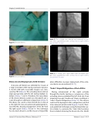
Figure 4. Extracted device.
Figure 5. Extracted device.
Dilator-Assisted Deployment of ASD Occluder
A 62-year-old female was admitted for closure of a large secundum ASD causing exertional dyspnea. A 32-mm ASD with reasonable margins was mea- sured by TEE (Video 6). The occluding device did not align appropriately with the IAS during multiple at- tempts (Videos 7 and 8). Therefore, the long dilator of the device delivery system was introduced over a J-wire through an additional venipuncture (Video 9). The dilator was used to retain the left disc in the LA as the right disc was uncovered and pulled gently to- ward the RA, allowing engagement of the IAS from the right aspect (Videos 10 and 11). This technique has also been shown to aid the closure of large ASDs
Video 5. The occluder was released and remained in posi- tion. View supplemental video at http://dx.doi.org/10.12945/j. jshd.2016.005.16.vid.05.
Video 6. A 32-mm atrial septal defect with reasonable mar- gins. View supplemental video at http://dx.doi.org/10.12945/j. jshd.2016.005.16.vid.06.
when di culties in proper deployment of the occlu- sive device are encountered [10, 11].
“Cobra”-Shaped Dis guration of the Left Disc
During advancement of the septal occluder through the sheath, twisting or compression of the occluding device prohibited the left atrial disc from acquiring its proper “mushroom” shape, resulting in a “cobra”-shaped dis guration (Video 12). The devices could not be deployed in this con guration and had to be removed and discarded (Figures 4 and 5). Previ- ously published techniques using the Amplatzer sep- tal occluder to overcome this “cobra”-like formation have included retrieving the device into the sheath
Journal of Structural Heart Disease, February 2017
Volume 3, Issue 1:15-27
