
Page 26 - Journal of Structural Heart Disease Volume 3, Issue 1
P. 26
19
Original Scienti c Article
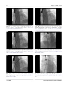
Video 7. The occluder failed to align appropriately with the interatrial septum. View supplemental video at http://dx.doi. org/10.12945/j.jshd.2016.005.16.vid.07.
Video 8. Another failed attempt to align the occluder with the interatrial septum. View supplemental video at http://dx.doi. org/10.12945/j.jshd.2016.005.16.vid.08.
Video 9. The dilator was introduced to facilitate deployment. View supplemental video at http://dx.doi.org/10.12945/j. jshd.2016.005.16.vid.09.
Video 10. The dilator retained the left disc in the left atrium, allowing engagement of the interatrial septum from the right. View supplemental video at http://dx.doi.org/10.12945/j. jshd.2016.005.16.vid.10.
Video 11. After successful deployment the dilator is with- drawn. View supplemental video at http://dx.doi.org/10.12945/j. jshd.2016.005.16.vid.11.
Video 12. “Cobra”-shape dis guration of the left disc. View sup- plemental video at http://dx.doi.org/10.12945/j.jshd.2016.005.16. vid.12.
Tal, R. et al.
Atrial Septal Defect Occlusion Challenges
