
Page 29 - Journal of Structural Heart Disease Volume 3, Issue 1
P. 29
Original Scienti c Article
22
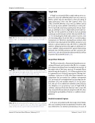
Video 19. The device assumed a normal position. View supple- mental video at http://dx.doi.org/10.12945/j.jshd.2016.005.16. vid.19.
Video 20. Secundum atrial septal defect near the superior vena cava opening into the right atrium. View supplemental video at http://dx.doi.org/10.12945/j.jshd.2016.005.16.vid.20.
Video 21. Contrast injection in the superior vena cava con rmed no obstruction to superior vena cava ow. View supplemental video at http://dx.doi.org/10.12945/j.jshd.2016.005.16.vid.21.
“High” ASD
A "high"-positioned ASD, located in the postero-su- perior IAS, must be di erentiated from sinus venosus defect, which may be described as the unroo ng of right pulmonary veins into the superior vena cava (SVC) or the RA. Whereas sinus venosus defect cannot be closed by transcatheter intervention in the pres- ence of partially anomalous pulmonary venous drain- age, an attempt to close a high ASD can be made. Us- ing TEE, we encountered an ASD in close proximity/ continuity to the entrance of the SVC into the RA (Vid- eo 19). Occluding this ASD with a device carries a risk of restricting in ow from the SVC [21] and thrombus formation [22]. Therefore, during the procedure, after the device was deployed in the defect and prior to its release, contrast injection into the SVC in a steep left anterior oblique projection through an additional ve- nous catheter demonstrated the spatial relationship between the SVC-RA junction and the occlusive de- vice (Video 20) and showed unobstructed blood ow from the SVC to the RA (Video 21).
Large Chiari Network
The Chiari network is a fenestrated membrane con- sisting of threads and strands in the RA. It is a congen- ital remnant resulting from incomplete resorption of the right valve of the sinus venosus. Prominent Chiari network may be found in 2–3% of the population, but it is generally not of clinical importance. During tran- scatheter occlusion of ASD, the Chiari network can complicate the procedure by catheter entrapment [23], proximal disc entanglement and inadequate de- ployment [24], and residual shunt [25]. In our situa- tion, the use of an additional catheter in the SVC was also helpful as in our previous case of “high” ASD. A catheter advanced from the femoral vein to the SVC may retain the Chiari network away from the IAS, thus lowering the probability of device or catheter entan- glement.
Double Interatrial Septum
A 37-year-old patient with end-stage renal failure, who was awaiting renal transplantation from his wife, was referred for an elective percutaneous closure of
Journal of Structural Heart Disease, February 2017
Volume 3, Issue 1:15-27
